An analysis of diet-induced obesity using MouseMapper—a suite of foundation-model-based deep-learning algorithms—identifies structural alterations of the infraorbital branch of the trigeminal ganglia.
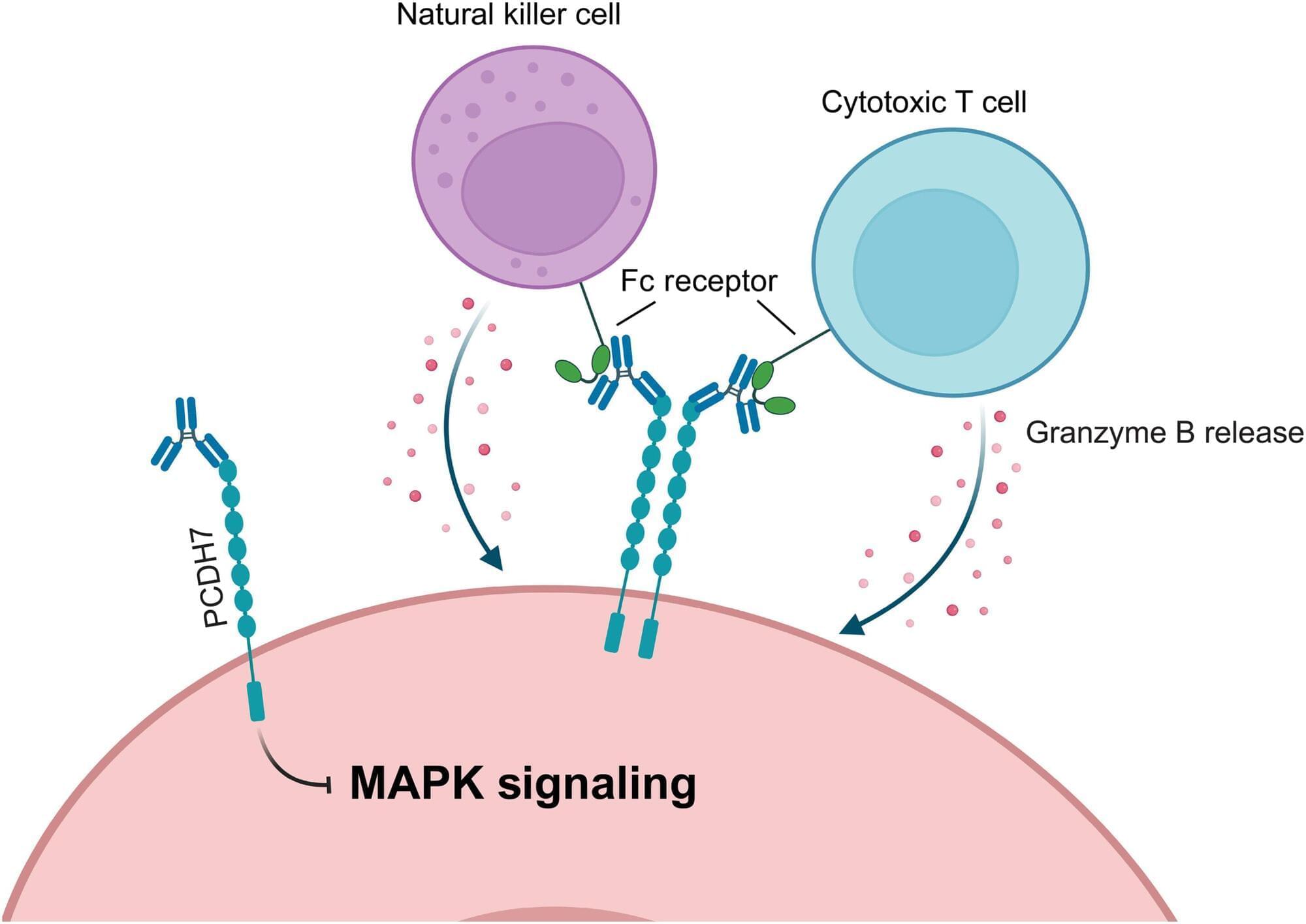
Scientists found that blocking a protein best known for its role in asthma enhances cancer immunotherapy in preclinical models.
On May 19, Dallas-based Colossal Biosciences, which last year made headlines when it effectively de-extincted the dire wolf, announced that it had hatched a flock of 26 live chicks from fully artificial eggs. The technology behind the breakthrough can be later applied to bring back the dodo and New Zealand’s giant, flightless moa—both on Colossal’s de-extinction ‘to do’ list…
…Designing an artificial shell is not easy because a natural shell is deceptively complex. Made principally of calcium carbonate arranged in a crystalline structure, a typical egg shell is no more than 0.4 mm thick, and covered with up to 17,000 tiny pores to allow for gas exchange with the ambient atmosphere—carbon dioxide out, and oxygen in. There are, too, a pair of slick inner membranes in the egg that perform another critical function, protecting the growing chick from invading bacteria. But those membranes have to be exceedingly thin…
…The egg Colossal invented was very different. The inner membranes were made of vanishingly thin silicon using a proprietary technology that Colossal is planning to patent. The shell itself was only about two-thirds of a shell—a titanium structure that resembles nothing so much as a soft-boiled-egg cup with its top missing, albeit with hundreds of hexagonal pores to allow for gas exchange. Once a few dozen of the titanium eggs were manufactured, Colossal gathered fertilized chicken eggs from an avian farm the company owns and operates and transported them to the lab. There, the scientists gently opened the top of the egg and transferred the yolk and the white and the tiny embryo onto the titanium egg cup and covered the cup with a transparent lid. The embryos were about three days past fertilization when they were transferred, meaning that they had 18 days remaining in their three-week incubation cycle.
‘We place the egg into an incubator that controls the environment,’ says Lambert. ‘We then collect visual images at periodic milestones to understand how development is progressing.’ When the incubation period was done, the chicks began ‘pipping,’ using their beaks to break through the membrane just the way an ordinary chick breaks through its shell. Eventually, the 26 chicks were moved to the same Texas farm from which their eggs were collected, where they can live out their five to 10 year lifespan.
The breakthrough could help bring giant birds back from extinction.
Aging human breast atlas reveals cancer susceptibility
The team used advanced imagining techniques to analyse breast tissue from more than 500 women aged 15 to 86 years old. The tissue included biopsies taken from women for non-cancer-related reasons.
Combining these images with details of the hormone receptors and immune cells present, as well as the tissue architecture, the researchers were able to map how breast tissue changes over time in unprecedented detail. Their findings point to reasons why breast cancer risk increases with age and why tumors in younger women differ biologically.
The author added: “Our map revealed that as women age, their breast tissue goes through major changes, with the most dramatic changes occurring at menopause. There are changes, too, during their twenties, possibly linked to pregnancy and childbirth, but these are far less pronounced.”
The map revealed that all types of cells become fewer in number and divide far less often. Milk-producing structures known as lobules shrink or disappear, while the ducts that that carry milk become relatively more common, with the supporting layer around them becoming thicker. Fat cells increase while blood vessels decrease.
Meanwhile, changes occur in the immune environment. Younger breasts have more B cells and active T cells, which helps them identify and kill cancer cells. As tissue ages, these types of cells decline in number, replaced by other types of immune cell that indicate a more inflammatory and potentially less protective immune environment. ScienceMission sciencenewshighlights.
The protein p53 is often called the guardian of the genome for its central role in preventing cancer. Yet paradoxically, it is also one of the most frequently mutated and dysfunctional proteins in human tumors.
A longstanding mystery has been why p53—unlike its closely related paralogs p63 and p73—is so prone to misfolding and forming toxic aggregates. A new study published in Communication Chemistry now provides a detailed molecular explanation for this vulnerability.
Led by researchers at the Federal University of Rio de Janeiro (UFRJ), the D’Or Institute for Research and Education (IDOR), the University of Campinas (Unicamp), and the Federal University of Tri ngulo Mineiro (UFTM), the team mapped the protein’s internal landscape at residue-level resolution using high-pressure NMR spectroscopy, fluorescence spectroscopy, and molecular dynamics simulations.
Scientists at the UCLA Health Jonsson Comprehensive Cancer Center have developed a new cytokine-armored CAR-T cell therapy that helps the immune system better attack aggressive brain tumors in mice while reducing dangerous side effects that have long limited immune-based treatments for glioblastoma, one of the deadliest and most treatment-resistant brain cancers.
The therapy works by reprogramming CAR-T cells to release immune-stimulating proteins, called IL-12 and DR-18, that activate the body’s own immune system, strengthening the overall anti-cancer response. In mouse models, the approach improved tumor control, including against cancers made up of mixed cell populations that often escape therapies.
Researchers also found that pairing the treatment with a second CAR-T strategy targeting VEGF, a protein that drives abnormal blood vessel growth and contributes to swelling in glioblastoma, helped reduce side effects while preserving strong anti-tumor activity.
“We are in a time of circulating flesh.”
Stelarc said that to me 13 years ago. In 2026, it reads less like art criticism and more like a status report.
He had grown an ear on his arm. He had hung himself from hooks 25 times. He had let strangers on the internet choreograph his muscles through electrical stimulation, his body remote-controlled across continents.
Most people called it spectacle. I think it was inquiry.
Because long before deepfakes, before voice cloning, before AI agents wearing our faces, was already asking the question we now cannot avoid:
Where does the body end and the network begin?
Many plastic products are designed to be used only once, yet the material itself lasts for years. But a new strategy is addressing this problem by creating products that self-destruct on command, known as living plastics. These materials incorporate activatable, plastic-degrading microbes alongside the polymers. One team reporting in ACS Applied Polymer Materials used two bacterial strains that worked together and completely broke down the material within just six days, without making microplastics.
Why scientists are rethinking plastics Zhuojun Dai, a corresponding author on the paper, explains that “the realization that traditional plastics persist for centuries, while many applications, like packaging, are short-lived, led us to ask: Could we build degradation directly into the material’s life cycle?”
Many microbes can break long polymeric chains into smaller pieces using enzymes. Because plastics are polymers, these enzymes or the microbes that make them could be incorporated into living plastics.
Breakthrough in brain medicine: a new way to deliver CBD!
Cannabidiol (CBD) has incredible potential to fight brain inflammation, but it has always faced a major roadblock: it struggles to dissolve and cross the blood-brain barrier. Researchers have just developed an ingenious solution using glucose-coated nanoparticles to get CBD exactly where it needs to go.
Here’s why it’s a game-changer: 🔬 Sneaky Delivery: The glucose coating helps the particles “hitch a ride” on the brain’s natural glucose transporters, successfully smuggling the CBD across the blood-brain barrier. 🎯 Smart Release: Once inside the brain, the nanoparticles target immune cells (microglia) and only release the CBD when they detect the chemical stress of active inflammation. 🐁 Promising Results: In mouse models of Parkinson’s disease and depression, this new delivery method drastically reduced inflammation, protected neurons, and improved behavioral recovery compared to standard CBD.
This targeted approach could be a massive step forward in treating chronic neuroinflammatory diseases! 🧬✨
Studty.
Glucose-coated nanoparticles carry CBD across the blood-brain barrier, trigger release in inflamed tissue, and reduce neuroinflammatory signs in mice.