The first evidence may not appear where we used to look.
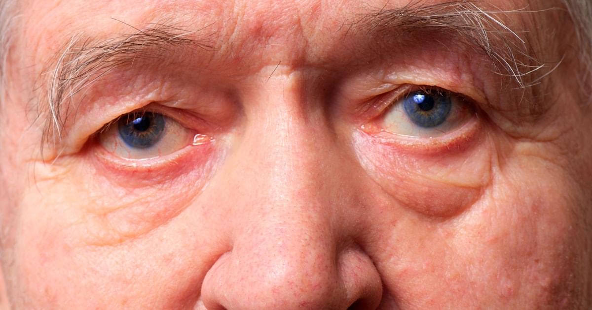
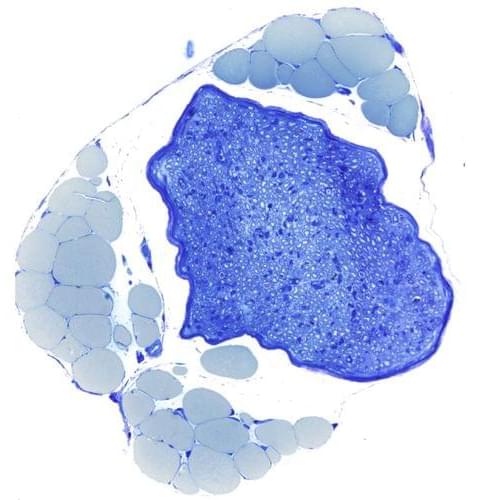
Damage to the body’s peripheral nerves can cause pain and movement disorders. Researchers at Leipzig University have recently investigated how damaged nerves can regenerate better. They found that fat tissue strongly supports the Schwann cells needed for repair during the healing process. The results were published in the renowned journal “Cell Metabolism”
Our bodies are transversed by millions of nerve fibres that transmit information. This allows us to do things like control muscles and perceive sensory impressions. Peripheral nerves, like those in our arms and legs, are often damaged by acute injuries, for example, in accidents. As a result, those affected suffer from loss of muscle strength and sensory problems such as numbness. Peripheral nerves do have a strong regenerative potential, but complete recovery of nerve function is still rare for reasons that are not yet fully understood.
When a nerve is crushed or severed, the individual nerve fibres affected by the damage initially die. In principle, they have the ability to grow back and regenerate completely. This depends on the Schwann cells that surround the nerve fibres. These cells do not die after nerve damage, but instead are responsible for coordinating the breakdown and regrowth of nerve fibres in their original areas. Schwann cells therefore play a key role in the repair process. It was previously unknown how these cells cope with the enormous metabolic load associated with the breakdown and rebuilding of nerve tissue. Researchers at the University of Leipzig Medical Center have now discovered that Schwann cells receive crucial support with nerve repair from the fat tissue that surrounds nerves in the body. Using genetically modified mice, they have shown that the chemical messenger leptin plays a key role in this process.
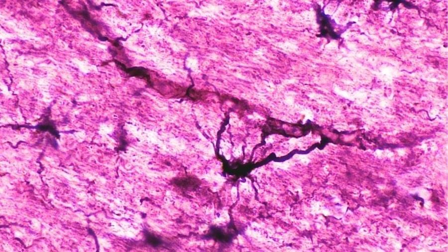
The severe and aggressive autoimmune disease known as neuromyelitis optica (NMO) just met a new match.
Without treatment, NMO can lead to serious disability, as rogue antibodies (AQP4-IgG) destroy the astrocyte support cells in the brain and spinal cord.
Therapies do exist to manage the condition, but they’re expensive, not always effective, and come with risks of their own – and relapses are common.

1 Center for Cutaneous Biology and Immunology Research, Department of Dermatology, Henry Ford Health, Detroit, Michigan, USA.
2Department of Plastic Surgery, Henry Ford Health, Detroit, Michigan, USA.
3Center for Bioinformatics, Department of Public Health Sciences, Henry Ford Health, Detroit, Michigan, USA.
The Neuroscience of Patriotism: From “Them” to “Us”
On the 4th of July, patriotism is often framed as flags, fireworks, and national pride. But neuroscience suggests something deeper: shared national identity can actually reshape how the brain processes other people.
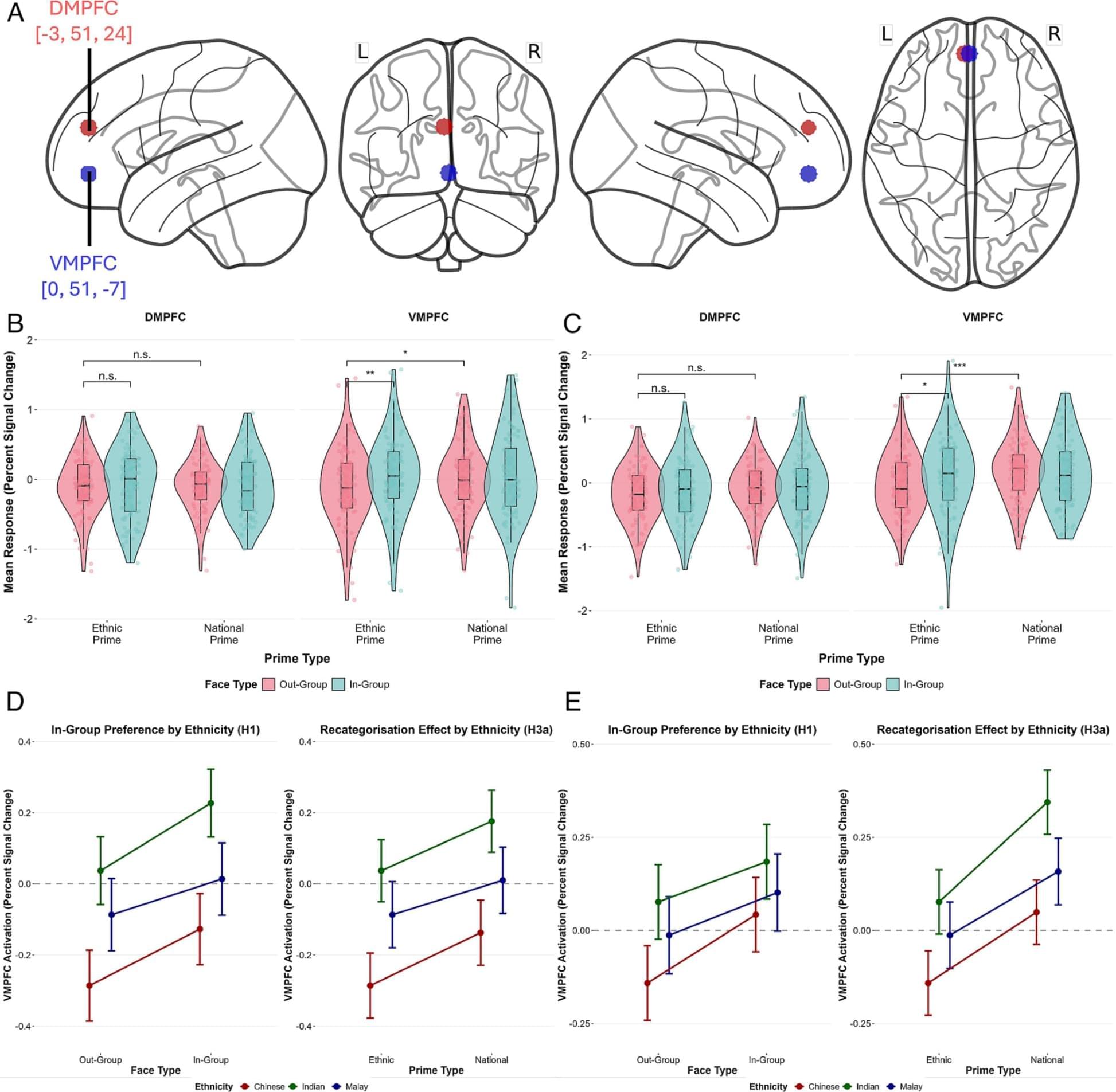
A 2026 fMRI study published in PNAS found that when people were briefly reminded of a shared national identity, their brains began responding more inclusively to faces from ethnic outgroups.
The key region was the ventromedial prefrontal cortex, an area involved in self-referential and social processing. Under ethnic identity cues, this region responded more strongly to ethnic ingroup faces. But under national identity cues, it showed increased engagement toward ethnic outgroup faces too.
In other words, the brain’s sense of “us” is flexible.
The study did not show that ethnic identity disappears. Instead, it suggests that shared identity can partially expand the boundary of belonging while still allowing subgroup identities to remain intact.
That may be one of the healthier forms of patriotism: not “my group above yours,” but “a larger we.”
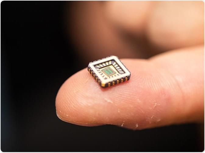
Artificial brain cells could now be implanted in the brain to repair the damage caused by chronic diseases, such as Alzheimer’s disease and other neurodegenerative conditions, thanks to a team of scientists who created bionic neurons that work like the real thing.
A team of scientists at the University of Bath created artificial neurons that could potentially help overcome paralysis, connect minds to machines, and restore failing brain circuits.
The new technology can help patients who have degenerative diseases affecting the brain.

While RNS and DBS are brain implants on the market with on and off label usages, there is also a class of brain implant devices which are purely in the clinical research realm. These brain machine interfaces use microelectrodes which record cellular level data and allow machine learning algorithms to control computer cursors and robotic arms. The first demonstration of this type of device’s efficacy was in non-human primates by the seminal work of Drs. Dawn Taylor, Andrew Scwartz, and colleagues.41 The microelectrode array, the ‘Utah array,’ was created in Salt Lake City, Utah, by the pioneering implant company, now called Blackrock Neurotech (Salt Lake City, Utah). This 4 × 4 mm array resembles a pin cushion that gets impacted into the cortical tissue with a precise pressurized insertion device (Figure 3). The adaptive-learning algorithm was engineered to sense neuronal firing patterns from the brain tissue and then uses those signals to control a device such as a computer cursor or robotic arm based on these patterns. The concept of ‘decoding neural data’ using machine learning is the foundation of BMIs and came from work by Dr. Schwartz and his mentor Dr. Apostolos Georgopoulos. Amazingly, animals and patients can adapt their own neural activity in motor cortex or parietal cortex through training an adaptive computer algorithm to learn the patient’s brain signals related to the intention to move, and then moving a robotic arm with varying degrees of freedom accordingly. Here AI is the computer model that trains on neural activity related to the desired output such as a robotic arm movement. This model learns a ‘transform function’ which it uses to predict when and how the patient wants to move the robotic arm in a future planned movement. Once trained, the patient can control a machine using the brain implant with their mind. The machine is effectively “mind-reading” via the learned transfer function. This concept opens the door to treating patients who are tetraplegic or otherwise locked-in and unable to communicate or interact with the world. It also leads to some interesting privacy issues such as, should and could there be controls in place for the computer not to read certain types of neural signals?
The first use of brain implants to treat such patients was led by Drs. John Donoghue, Leigh Hochberg, and their team at Brown University and Massachusetts General Hospital, via the BrainGate clinical trials.42,43 The BrainGate2 clinical trial (NCT00912041) is currently active and recruiting patients with tetraplegia from amyotrophic lateral sclerosis or spinal cord injury. These patients have a Blackrock NeuroPort electrode-based BCI device implanted into the motor cortex or other cortical areas. Patients use their brain activity to train a machine learning algorithm to then control an assistive device. While these clinical trials are certainly tailored to the individual patient, these trials help researchers develop better control algorithms for other BCI applications and helps researchers gain insights into how the human brain works, which they otherwise would not be able to learn. For example, in a study with stroke patients at Washington University in St. Louis, it was noted that patients could control the limb ipsilateral to a control device in motor cortex, when generally we do not think about possible ipsilateral limb control capabilities of motor cortex.44 Note that the Blackrock NeuroPort electrode (which is the human version of the Utah array) is not fully implanted. It requires a head-mounted pedestal to transfer data and that piece is exposed outside the skin which may carry a higher risk of infection than a fully implanted device.45 Neuralink’s (Fremont, California) N1 Chip mentioned above, is fully implantable and has 1,024 electrodes. Several patients with tetraplegia or tetraparesis have been implanted with this research device in the ongoing PRIME clinical trial (NCT06429735). Paradromics (Austin, Texas) has the Connexus BCI interface that is also fully implantable and supports 1,600+ channels of data, again supporting AI models that require large amounts of data and has also been implanted in humans. Precision (New York City, New York) has a thin seven-layer film designed to capture data at the level of LFPs (NCT05182437) and is designed to treat epilepsy. It is also fully implantable with a battery in the chest and can capture wave phenomena on the brain and has been implanted in several patients. Finally, Synchron (Brooklyn, New York) has created the Stentrode, which is a device with electrodes mounted on a stent that is then implanted in a cerebral vessel near motor cortex. The device records cortical neural activity that is rich enough to run an AI algorithm to control a touchscreen device. The potential advantage here is perhaps a lower rate of infection by being intravascular, as opposed to the immune sheltered environment of the brain. The SWITCH trial (NCT 03834587) enrolled five patients with results pending.
Aside from motor control, speech prostheses designed for communication have also emerged. Here the concept is to decode speech directly from speech-related motor areas including ventral sensorimotor cortex and midprecentral gyrus using a brain implant.46 Patients most appropriate have motor paralysis causing dysarthria or anarthria, which is the total inability to produce speech. This could be a result of stroke or amyolateral sclerosis. First demonstrations of speech decoding came from the lab of Edward Chang, MD, followed by others.46 This does require that the patient’s ability to understand speech is intact. The control signal is generated usually by imagining the speech. Most recent iterations involve a patient having an avatar perform realistic facial movements as well as generate something similar to the patient’s voice.47 Here you can imagine that if the decoding is accurate, any words the patient imagines would be projected, which may compromise patient privacy to some degree.

From the air we breathe to the food we eat, we are constantly exposed to thousands of chemicals—yet how these exposures affect our health has remained surprisingly difficult to understand. A new study led by researchers at the CeMM Research Center for Molecular Medicine of the Austrian Academy of Sciences and the Ludwig Boltzmann Institute for Network Medicine at the University of Vienna, published in Nature Communications, offers a unifying view: Diverse substances can disrupt the same biological systems and thereby contribute to disease risk in predictable ways.
Environmental pollution is estimated to contribute to around one in six deaths worldwide, but scientists have long struggled to connect specific exposures to specific diseases. One reason is the sheer complexity of the “exposome” —the totality of all environmental influences a person encounters over a lifetime. Traditionally, chemicals have been grouped by their structure or origin, but this says little about what they actually do inside the body. Two nearly identical molecules can have completely different effects, while entirely unrelated substances may trigger the same illness. This has made it difficult to move from observation to understanding.
A new study, led by Jörg Menche, CeMM adjunct PI and director of the Ludwig Boltzmann Institute for Network Medicine, and first authored by former Ph.D. student at CeMM and LBI NetMed (now a postdoc at Harvard Medical School) Salvo Danilo Lombardo, takes a different route: Instead of asking what chemicals look like, the researchers asked what they do. They compiled nearly 10,000 environmental exposures, ranging from pollutants and food components to medications, and mapped how each affects human genes. The result is a large-scale network that links exposures based on shared biological effects.