Researchers have developed a revolutionary biosensor using terahertz (THz) waves that can detect skin cancer with exceptional sensitivity, potentially paving the way for earlier and easier diagnoses. Published in IEEE Transactions on Biomedical Engineering, the study presents a significant advancement in early cancer detection, thanks to the collaboration of multidisciplinary teams from Queen Mary University of London and the University of Glasgow.
“Traditional methods for detecting skin cancer often involve expensive, time-consuming, CT, PET scans and invasive higher frequencies technologies,” explains Dr. Shohreh Nourinovin, Postdoctoral Research Associate at Queen Mary’s School of Electronic Engineering and Computer Science, and the study’s first author. “Our biosensor offers a non-invasive and highly efficient solution, leveraging the unique properties of THz waves—a type of radiation with lower energy than X-rays, thus safe for humans—to detect subtle changes in cell characteristics.”
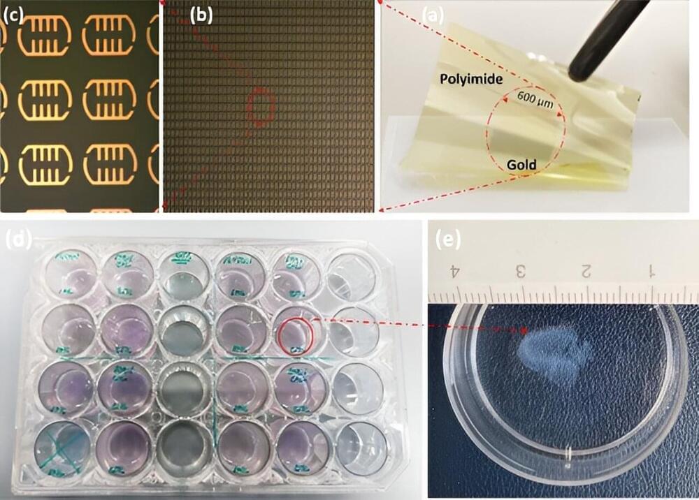
The key innovation lies in the biosensor’s design. Featuring tiny, asymmetric resonators on a flexible substrate, it can detect subtle changes in the properties of cells. Unlike traditional methods that rely solely on refractive index, this device analyzes a combination of parameters, including resonance frequency, transmission magnitude, and a value called “full width at half maximum” (FWHM). This comprehensive approach provides a richer picture of the tissue, allowing for more accurate differentiation between healthy and cancerous cells and to measure malignancy degree of the tissue.