Toggle light / dark theme

Mechanical load inhibits cancer growth in mouse and human hearts

The heart’s constant beating may actively suppress tumor growth in cardiac tissues, a new Science study reports. This is because cellular pathways in these tissues alter gene regulation in cancer cells to keep them from proliferating.

The findings shed light on the role of mechanical forces in protecting the heart from cancer and may pave the way to new cancer therapies based on mechanical stimulation.


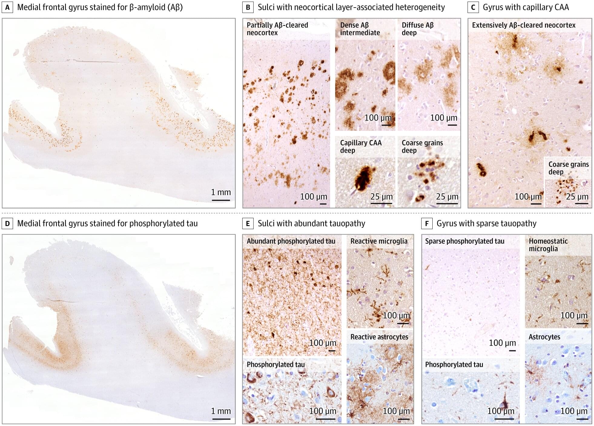
The heart rarely develops cancer, and, at the same time, it lacks regenerative capacity, as cardiomyocytes stop proliferating after birth. This suggests that mechanisms limiting cardiac regeneration may also protect against cancer. In this work, we investigated the role of mechanical load and used in vivo cancer models and ex vivo engineered heart tissues to show that mechanical load reduces cancer cell proliferation in the myocardium. Spatial transcriptomics of human cardiac metastases revealed decreased histone methylation and chromatin compaction. These changes affect chromatin accessibility at proliferation-related loci, with Nesprin-2 identified as a key mechanosensor. Our results uncover how mechanical forces protect the heart from cancer and suggest potential strategies for cancer therapy based on mechanical stimulation.

Performance of a large language model on the reasoning tasks of a physician

What if every scientific paper you read was just the “highlight reel” of a much longer, messier, and more complicated movie? You see the breakthrough, but you never see the hundreds of hours of footage showing what didn’t work.

Ultimately, the ARA marks a shift toward a future where “The Last Human-Written Paper” isn’t the end of science, but the beginning of a much deeper, machine-readable conversation.

However, this shift toward radical transparency comes with its own set of hurdles. While ARAs make AI agents more efficient, the study found a “prior-run box” effect where seeing a human’s past failures actually limited an AI’s ability to think outside the box and find creative new solutions. There is also a significant cultural and technical gap to bridge: the system relies on researchers being willing to expose their “messy” unfinished work, and even with better data, the jump in actual experiment reproduction was relatively modest. Furthermore, the reliance on “compilers” to translate old papers into this new format risks baking in errors or “hallucinations” if the original source was vague, proving that while machine-readable data is powerful, it isn’t a magic fix for the inherent complexities of scientific discovery.


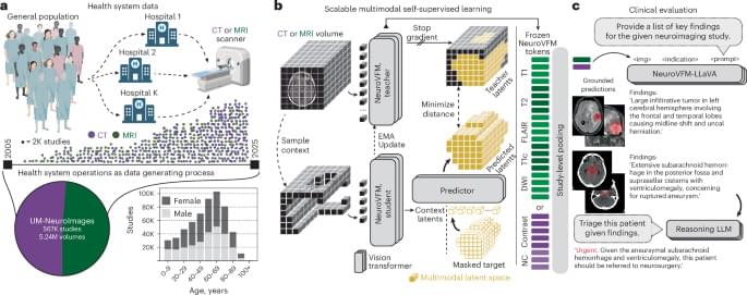
We systematically evaluated the medical reasoning abilities of an LLM across six diverse experiments, comparing the model with hundreds of expert physicians. Overall, the model outperformed physicians across experiments, including in cases utilizing real and unstructured clinical data taken directly from the health record in an emergency department. These diagnostic touchpoints mirror the high-stakes decisions taken in emergency medicine departments, where nurses and clinicians make time-sensitive choices with limited information. Our results showed that humans, GPT-4o, and o1 all improved their diagnostic abilities as more information was available; o1 outperformed humans at multiple touchpoints, with the widest gap at initial ER triage, where there is the least information available.

The rapid pace of improvement in LLMs has substantial implications for the science and practice of clinical medicine. Although applying AI to assist with clinical decision support is sometimes viewed as a high-risk endeavor (22, 23), greater use of these tools might serve to mitigate the human and financial costs of diagnostic error, delay, and lack of access (24, 25). Our findings suggest the urgent need for prospective trials to evaluate these technologies in real-world patient care settings and for health care systems to prepare for investments for computing infrastructure and design for clinician-AI interaction that can facilitate the safe integration of AI tools into patient-care workflows. This includes the development of robust monitoring frameworks to oversee the broader implementation of AI clinical decision support systems (22), monitoring not just final diagnostic accuracy but other metrics crucial for successful deployment, including safety, efficiency, and cost.

We emphasize that our study addresses only text-based performance for both humans and machines; clinical medicine is multifaceted and awash with nontext inputs, including auditory (such as the patient’s level of distress) and visual information (for example, interpretation of medical imaging studies) that clinicians routinely use. Existing studies suggest that current foundation models are more limited in reasoning over nontext inputs (26, 27); future work is needed to assess how humans and machines may effectively collaborate (28) in use of nontext signals. This requires new benchmarks, trials, and technological solutions to more faithfully measure clinical encounters. Existing investment in increasingly pervasive ambient AI scribes and other passive monitoring technologies holds promise to serve as the basis for such investigations.

Metastatic cancer detection and management with artificial intelligence and augmented reality (Review)

Metastatic cancer remains a significant global health challenge, contributing to the majority of cancer-related mortality due to late detection, therapeutic resistance and the complexity of disseminated disease. Recent advances in artificial intelligence (AI) and augmented reality (AR) are transforming the landscape of metastatic cancer detection and management. AI-driven tools, including radiomics, deep learning models, and predictive analytics, enhance early identification of metastatic lesions, improve diagnostic accuracy, and support personalized treatment strategies by integrating multimodal clinical, imaging and molecular data. At the same time, AR technologies are increasingly applied in image-guided surgery, real-time tumor visualization and patient education, enabling more precise interventions and improved clinical decision-making.

Common asthma drug may turn off tumor ‘switch’ tied to immunotherapy resistance

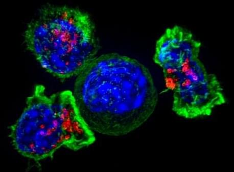
A drug widely used to treat asthma and allergies may also help fight aggressive cancers, reports a new Northwestern Medicine study that uncovered how tumors hijack common white blood cells to evade immunotherapy.

The findings in mice and human tissues point to a practical, new way to improve treatment for tough tumors, such as triple-negative breast cancer, where immunotherapy often fails.

The study is published in Nature Cancer.

Dietary fats shape pancreatic cancer risk via ferroptosis

For decades, the relationship between fat and cancer has been treated as a question of quantity: Eat less fat, reduce your risk of developing cancer. But new research published April 29 in Cancer Discovery shows that for pancreatic cancer, the type of fat you consume matters more than the amount.

“It’s really the type of fat that you’re consuming, not just total fat content,” says Christian Felipe Ruiz, Ph.D., an associate research scientist in YSM’s Department of Genetics and lead author of the study. “Depending on the type of fat that you consume, it can go completely different ways. We found that some fats promote cancer, as we would expect, while other fats are really good at suppressing cancer.”

One fat in particular—oleic acid, the primary fatty acid in olive oil—may be accelerating tumor growth in ways scientists never anticipated. The result was surprising given oleic acid’s reputation in medicine. “It’s traditionally been considered a healthy type of fat for cardiovascular health,” Ruiz says.

Age does not appear to drive cardiovascular risk in pregnancy

Underlying cardiovascular risk, rather than older age, drives complications such as venous thromboembolism, cardiomyopathy and heart failure during pregnancy, according to new Weill Cornell Medicine research. The findings may encourage doctors to more actively address cardiovascular health in patients before they become pregnant.

The study, published in Nature Communications, suggests that instead of pregnancy becoming inherently riskier as people get older, it amplifies a person’s baseline cardiovascular risk, regardless of age.

“Pregnancy seems to be a uniform stress test, so to speak,” said the study’s lead author, Dr. Hooman Kamel, vice chair of clinical research and chief of neurocritical care in the Department of Neurology and the Helen and Albert Moon Professor of Neurology at Weill Cornell Medicine.

A new way to recharge aging muscle stem cells by restoring a key metabolic component

Losing muscle strength is a natural part of aging. At the core of this decline is a drop in the number of muscle stem cells (MuSCs), the specialized cells responsible for maintaining and regenerating muscle tissue throughout our lives. Loss of muscle strength can severely affect mobility, increasing the risk of falls, fractures and, most importantly, the loss of independence.

Published in Nature Aging, a recent study took a crucial first step toward restoring stem cell function in aging muscles—gaining a clearer understanding of how metabolism changes when stem cells are activated and how these critical processes weaken with age.

The researchers’ investigation led them to glutamine metabolism, the process by which cells use the amino acid glutamine to support essential functions. They found that for MuSCs, glutamine is more than just a nutrient. It provides the raw material needed to produce fatty acids that help cells grow, divide, and repair damaged muscles.

Bioengineers condense protein engineering and testing to a single day

Proteins are critical to life—and to industry. There are countless proteins that could be engineered to treat and even cure serious diseases and cellular dysfunctions. Industrial applications are similarly promising, with proteins increasingly used as enzymes in food manufacturing and in consumer detergents.

While AI can help suggest improvements, each novel protein must still be created in the real world and tested for performance. It is a labor-intensive process that involves constructing the DNA instructions for each protein in yeast or bacteria and growing individual clones for protein production and testing. This can take many days for a single protein of interest and even longer if the protein needs to be tested in mammalian cells, a process that requires retrieving DNA from microbes for transfer to the mammalian cells.

In a new paper, Michael Z. Lin, a professor of neurobiology and of bioengineering in the schools of Engineering and Medicine, and graduate students, Yan Wu in bioengineering and Pengli Wang in chemical engineering, say they have condensed the time-intensive protein building and testing process to just 24 hours.

/* */