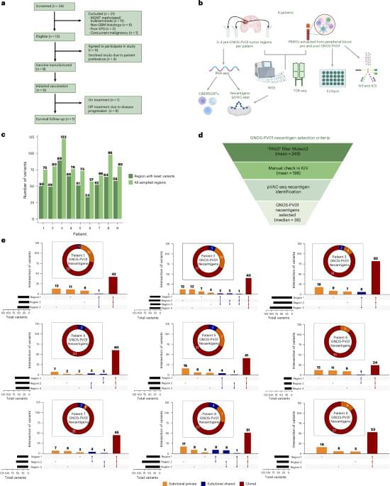
Toggle light / dark theme

A new atlas could help guide researchers studying neurological disease

Functioning brain cells need a functioning system for picking up the trash and sorting the recycling. But when the cellular sanitation machines responsible for those tasks, called lysosomes, break down or get overwhelmed, it can increase the risk of Alzheimer’s, Parkinson’s, and other neurological disorders.

“Lysosomal function is essential for brain health, and mutations in lysosomal genes are risk factors for neurodegenerative diseases,” said Monther Abu-Remaileh, a Wu Tsai Neuro affiliate and an assistant professor of chemical engineering in the Stanford School of Engineering and an assistant professor of genetics in the Stanford School of Medicine.

The trouble is, scientists aren’t sure exactly how lysosomes do their work, what’s going wrong with lysosomes that leads to neurodegeneration—or even in which cell types neurodegenerative disease begins. There might even be other lysosomal disorders yet to be discovered.

Growth chambers could enable reproducible plant-microbe data across continents

Harnessing the power of artificial intelligence to study plant microbiomes—communities of microbes living in and around plants—could help improve soil health, boost crop yields, and restore degraded lands. But there’s a catch: AI needs massive amounts of reliable data to learn from, and that kind of consistent information about plant-microbe interactions has been hard to come by.

In a new paper in PLOS Biology, researchers in the Biosciences Area at Lawrence Berkeley National Laboratory (Berkeley Lab) led an international consortium of scientists to study whether small plastic growth chambers called EcoFABs could help solve this problem.

Building on their previous work with microbe-free plants, the scientists used the Berkeley Lab-developed devices to run identical plant–microbe experiments across labs on three continents and got matching results. The breakthrough shows that EcoFABs can remove one of the biggest barriers in microbiome research: the difficulty of reproducing experiments in different places.

Molecular surgery: ‘Deleting’ a single atom from a molecule

Inserting, removing or swapping individual atoms from the core of a molecule is a long-standing challenge in chemistry. This process, called skeletal editing, can dramatically speed up drug discovery or be applied for upcycling of plastics. Consequently, the field is witnessing a surge of interest spanning from fundamental chemical research to applications in the pharmaceutical industry.

A group of researchers have now extended the scope of skeletal editing to the scale of just a single molecule. Such a level of precision in skeletal editing is unprecedented, and this may open a new route to obtain elusive molecules.

The team of researchers are active at Chalmers University of Technology, Sweden; IBM Research Europe—Zurich, Switzerland; and CiQUS at the University of Santiago de Compostela, Spain. In a recent article published in the Journal of the American Chemical Society, they demonstrate how, in a controlled manner, they can selectively remove a single oxygen atom from an organic molecule using the sharp tip of a scanning probe microscope.

AI method advances customized enzyme design

Enzymes with specific functions are becoming increasingly important in industry, medicine and environmental protection. For example, they make it possible to synthesize chemicals in a more environmentally friendly way, produce active ingredients in a targeted manner or break down environmentally harmful substances.

Researchers from Gustav Oberdorfer’s working group at the Institute of Biochemistry at Graz University of Technology (TU Graz), together with colleagues from the University of Graz, have now published a study in Nature describing a new method for the design of customized enzymes.

The technology called Riff-Diff (Rotamer Inverted Fragment Finder–Diffusion) makes it possible to accurately and efficiently build the protein structure specifically around the active center instead of searching for a suitable structure from existing databases. The resulting enzymes are not only significantly more active than previous artificial enzymes, but also more stable.

Neuropsychiatric symptoms in cognitive decline and Alzheimer’s disease: biomarker discovery using plasma proteomics

Placental toxicology progress!

Commonly used in vitro and in vivo placental models capture key placental functions and toxicity mechanisms, but have significant limitations.

The physiological relevance of placental models varies, with a general hierarchy of simple in vitro complex in vitro/ organ-on-chip in vivo, but species-of origin considerations may alter their relevance to human physiology.

Cellular, rodent, human, and computational modeling systems provide insights into placental transport, physiology, and toxicology linked to maternal–fetal health.

Recent advances in 3D culture and microfluidic technologies offer more physiologically relevant models for studying the placenta.

Mathematical modeling approaches can integrate mechanistic physiological data and exposure assessments to define key toxicokinetic parameters.

Environmental chemical concentrations and omic data obtained from placental tissues can link toxicant influences on placental function to adverse birth outcomes.

New Study Links Altered Cellular States to Brain Structure

Researchers at the Icahn School of Medicine at Mount Sinai have characterized how cellular senescence—a biological process in which aging cells change how they function—is associated with human brain structure in both development and late life. The study, published January 22 in Cell, provides new insight into how molecular signatures of cellular senescence that are present during development and aging mirror those associated with brain volume and cortical organization.

Understanding brain structure is a central challenge in neuroscience. Although brain structure changes throughout life and is linked to both aging and neurodegenerative conditions such as Parkinson’s and Alzheimer’s diseases, the underlying molecular processes involved—including cellular senescence—are not defined. Cellular senescence is commonly defined as a state characterized by permanent cell cycle arrest in the absence of cell death, in which cells have altered function. While cellular senescence has been implicated in aging and disease, its role in shaping human brain structure—both during development and aging—has remained unclear.

“This is the first study to directly link senescence-related molecular networks in living human brain tissue to measurable differences in brain structure within the same individuals,” said Noam Beckmann, PhD, Director of Data Sciences and founding member for the Mount Sinai Clinical Intelligence Center, Assistant Professor of Artificial Intelligence and Human Health, and co-senior author of the paper. “By identifying molecular pathways that are engaged in both brain structure development and aging, our work highlights senescence as a fundamental biological feature of brain aging and neurodegenerative disease and helps prioritize targets for future experimental research aimed at protecting brain health.”

Dried blood spot biomarker test for the detection of Alzheimer’s disease

The researchers tested a new method for detecting Alzheimer’s disease using a few drops of blood obtained from the fingertip and then dried on a card. This process was used to find proteins linked to Alzheimer’s disease and other brain changes in the 337 participants.

The study found that levels of p-tau217 in finger-prick samples closely matched results from standard blood tests and were able to identify Alzheimer’s disease-related changes in spinal fluid with an accuracy of 86 per cent. Two other markers, glial fibrillary acidic protein (GFAP) and neurofilament light (NfL), were also successfully measured and showed strong agreement with traditional tests.

While not ready for clinical use, this breakthrough addresses critical barriers in Alzheimer’s research by enabling remote participation in studies, clinical trial recruitment and monitoring, broader population sampling for epidemiological research, and inclusion of underrepresented communities and regions with limited healthcare infrastructure.

The findings suggest that this simple technique could make large-scale studies and remote testing possible, including for people with Down syndrome, who face a higher risk of Alzheimer’s disease and for other underserved populations. ScienceMission sciencenewshighlights.


A groundbreaking international study has demonstrated that Alzheimer’s disease biomarkers can be accurately detected using simple finger-prick blood samples that can be collected at home and mailed to laboratories without refrigeration or prior processing.

The research published in Nature Medicine. It represents the first large-scale validation of this accessible testing approach that removes geographic barriers and opens brain disease research to global populations without requiring specialised healthcare infrastructure.

Cellular senescence linked to brain structure changes across lifespan

Researchers at the Icahn School of Medicine at Mount Sinai have characterized how cellular senescence—a biological process in which aging cells change how they function—is associated with human brain structure in both development and late life.

The study, published in Cell, provides new insight into how molecular signatures of cellular senescence that are present during development and aging mirror those associated with brain volume and cortical organization.

Understanding brain structure is a central challenge in neuroscience. Although brain structure changes throughout life and is linked to both aging and neurodegenerative conditions such as Parkinson’s and Alzheimer’s diseases, the underlying molecular processes involved—including cellular senescence—are not defined.

Abstract: Infiltration of T cell acute lymphoblastic leukemia (ALL) into the meninges worsens prognosis

Ksenia Matlawska-Wasowska & team show T-cell leukemia exploits an inflammatory pathway to invade the brain’s protective layers, revealing a potential target for therapies aimed at preventing disease progression:

The image features GFP⁺ T-ALL leukemic infiltrates within whole-mount murine meningeal tissue. Credit: Wojciech Ornatowski.


1Department of Cell Developmental and Integrative Biology, University of Alabama at Birmingham, Birmingham, Alabama, USA.

2Department of Pediatrics, University of New Mexico, Albuquerque, New Mexico, USA.

3Department of Pediatrics, University of Alabama at Birmingham, Birmingham, Alabama, USA.

/* */