Few studies have directly examined the relationship between retinal structure and cognitive function in children. Provost et al. reported an inverse correlation of the dense retinal microvascular network with behavioral outcomes and sustained attention ability, which is consistent with our results66. However, no significant correlation between RNFL thickness and symptom severity of ADHD or EF performance has been reported. This indicates that although RNFL measurements may reflect certain neural characteristics of ADHD, they do not directly correlate with functional impairments67. Our ML model successfully stratified EF severity using retinal photograph analysis but did not achieve stratification of symptom severity as measured using the K-ARS. This corroborates the complexity of ADHD diagnosis and suggests that retinal biomarkers, although potentially valuable, should be considered complementary to conventional diagnostic tools, such as the DSM-5.
Nevertheless, the correlation between the retinal structure and EF deficits in ADHD supports its potential as a therapeutic biomarker. Notably, methylphenidate (MPH), a dopamine reuptake inhibitor, enhances various aspects related to attention in individuals with ADHD68, and significant differences in terms of parafoveal thickness were observed between the ADHD and TD groups, indicating a possible influence of MPH on retinal circulation69. Additionally, retinal thickness was positively correlated with the duration of MPH use, with greater retinal thickness in children undergoing treatment with MPH for 24 months70. Complementing these clinical findings, the animal study has shown that MPH exerts beneficial effects on the retina by reducing microgliosis, mitigating blood-retinal barrier dysfunction, and attenuating inflammatory responses71.
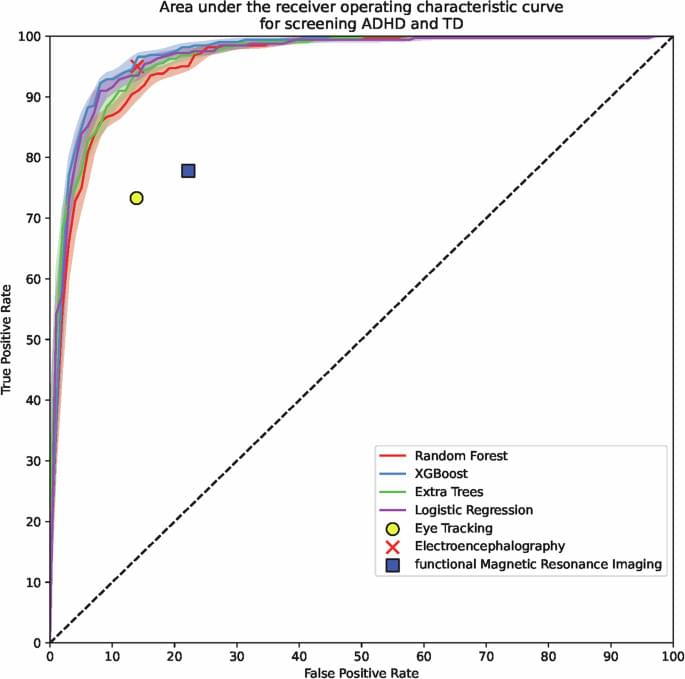
This study has some limitations. First, the dataset was derived from two hospitals in South Korea, potentially limiting the generalizability of our findings to broader populations. Future validation with external datasets encompassing diverse geographical and demographic settings is essential to confirm the global applicability of retinal biomarkers in ADHD screening and visual attention stratification. Nevertheless, our study provides robust preliminary evidence for the potential utility of retinal photographs in this context. Second, while retinal photographs were effective in identifying structural differences associated with ADHD, the two-dimensional nature of these images provides limited information. Although optical coherence tomography (OCT), incorporating three-dimensional imaging techniques, has been explored in ADHD and ASD populations, its results have been inconsistent25,72. Retinal photography, due to its rapidity and accessibility, may serve as a practical screening tool. On the one hand, advanced imaging modalities such as optical coherence tomography angiography (OCT-A) could complement retinal photography by capturing microvascular changes, presenting an alternative for further biomarker investigation. Third, our study focused on participants within a narrow age range (mean: approximately 9 years), which limits the generalizability of the findings to other developmental stages. Future research should investigate whether retinal biomarkers vary across age groups, considering the developmental trajectories of ADHD.