Toggle light / dark theme

Get the latest international news and world events from around the world.

Log in for authorized contributors

Radiation dose constraints for organs at risk in neuro-oncology; the European particle therapy network consensus

Case report: radiation-induced optic neuropathy following radiation therapy for a recurrent tuberculum sellae meningioma: a case report

🔗


Prior to the first surgery, the patient experienced progressive visual loss, resulting in a visual acuity of 20/200 in her right eye, and visual field loss in the temporally and lower 2 quadrants. Her vision was described as normal in the left eye prior to surgery. Visual function improved after surgery, and after 3 months visual acuity in the patient’s right eye was assessed to be 20/40. Despite the successful initial surgery, a tumor recurrence was detected 4 years later. At this time point, the patient had reduced vision and a progressive visual field loss in her right eye. After the second surgery, her visual acuity was 20/60 in her right eye and 20/22.5 in the left eye. The right visual field was substantially reduced, especially in the lower 2 quadrants.

Based on recurrent disease, high probability of microscopic residual tumor remnants, and progressive loss of visual function, the patient was offered postoperative radiation therapy delivered with photons. Radiation therapy was administered 4 months following the last surgical procedure, and a conservative fractionation of 1.8 Gy in 29 fractions, with a total dose of 52.2 Gy, was chosen to keep anterior visual pathway doses below what is considered safe (Table 1) according to the European Particle Therapy Network Consensus. These standards recommend doses below 55 Gy to 0.03 cm3 (D 0.03cc) to the chiasm and the optic nerves.1 For the chiasm, the maximum dose (D max) was 53.7 Gy and the mean dose (D mean) was 51.7 Gy. The right optic nerve had a D max of 53.3 Gy and a D mean of 48.8 Gy, whereas the left optic nerve had a D max of 52.3 Gy and a D mean of 30.5 Gy. Radiation therapy dose distribution is shown in Figure 3. After radiation therapy, the patient reported minor improvement in visual function. Unfortunately, 7 to 8 months later her visual function again deteriorated over only a few weeks. Both eyes were affected, and deterioration was most pronounced in her left eye, with only finger counting possible and nearly total visual field loss. In the right eye, visual acuity was 20/60, with a substantially reduced visual field, especially in the lower 2 quadrants.

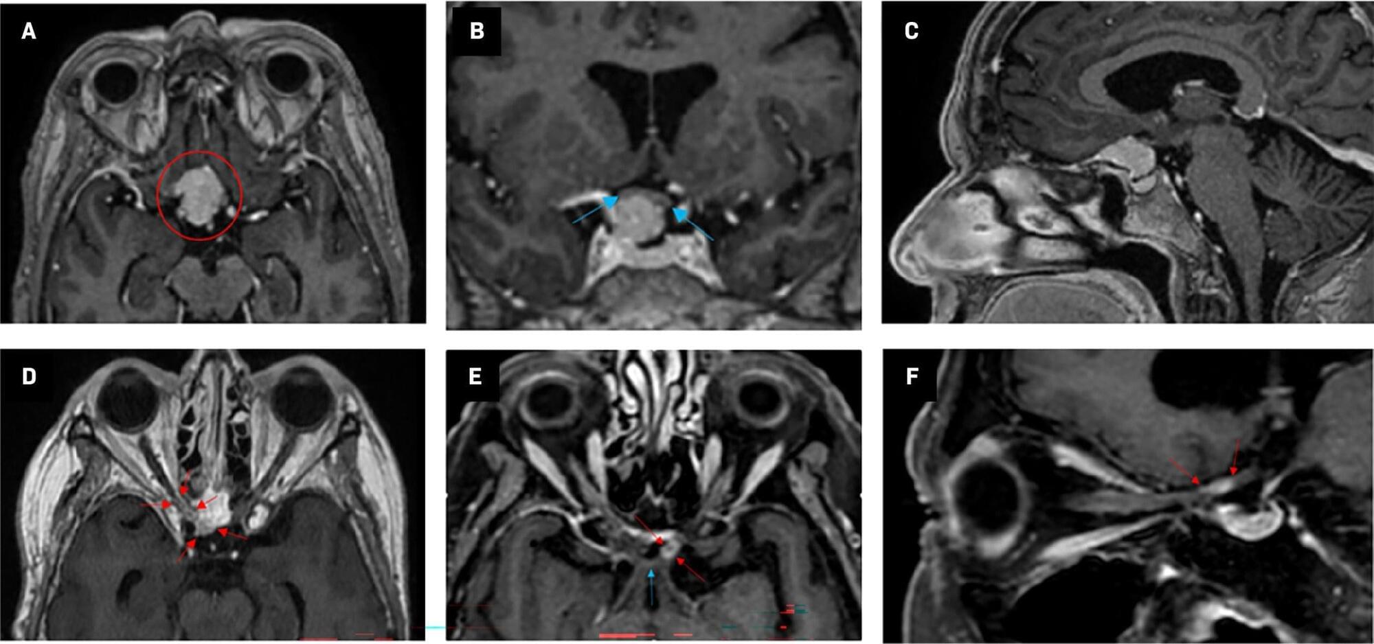
RION is primarily a diagnosis of exclusion, with tumor recurrence being the most important differential diagnosis. Tumor progression often results in a slower course of visual loss than RION.2 The patient’s vision loss was painless and rapid in onset, and radiological findings were bilateral and not consistent with neoplastic progression. Although MRI findings are nonspecific in RION-affected patients, neuro-ophthalmological examinations and clinical course/timing pointed to RION as the most likely cause of the patient’s visual loss. Systemic steroids in high doses were administered for 2 weeks before gradual tapering off, without any improvement in visual function. As a last effort to improve visual function, it was decided to try bevacizumab 7.5 mg/kg every 3 weeks. Treatment was well tolerated, with no reported side effects.

Radiation-Induced Optic Neuropathy Following Radiation Therapy for a Recurrent Tuberculum Sellae Meningioma: A Case Report

A new light-based imaging approach has produced an unprecedented chemical map of the Alzheimer’s brain.

Rice University researchers have produced what they describe as the first full, label-free molecular atlas of an Alzheimer’s brain in an animal model. In simple terms, they created a brain-wide “chemical map” that can help scientists study where the disease appears to take hold and how it spreads over time. Alzheimer’s is also a major public health threat, killing more people than breast cancer and prostate cancer combined.

Instead of focusing only on classic pathology markers, the team examined the brain’s underlying chemistry using a light-based imaging approach paired with machine learning. Their study, published in ACS Applied Materials and Interfaces, shows that Alzheimer’s-linked chemical shifts are patchy across the brain rather than uniform. It also suggests those shifts extend beyond amyloid plaques, the best-known feature of the disease.

A neurobiological perspective on prolonged grief disorder

The neurobiology of why some brains cannot move on from loss.


Prolonged grief disorder (PGD) is a psychiatric condition that describes individuals who experience persistent grief reactions characterized by preoccupation with the loss. This review provides an overview of the evidence on neurobiological processes associated with PGD. We propose that, although the neurobiological circuitry of PGD overlaps with that of anxiety and depression, it also involves neural responses that reflect the distinct symptom profiles of people with PGD. Specifically, while recruitment of cognitive control and salience networks is observed across common mental disorders, there is evidence that aberrant neural processes implicated in reward processes and appetitive functions are somewhat distinctive to PGD.

Chemists synthesize first stable copper metallocene complex, closing a 70-year gap

Almost half a century ago, a remarkable molecule called metallocene took center stage in chemistry, earning Geoffrey Wilkinson and Ernst Otto Fischer the Nobel Prize. These organic compounds, made of a transition metal “sandwiched” between two flat, ring-shaped organic layers, have since become an integral part of new-age polymers, materials, and pharmaceuticals.

In their recent work published in the Journal of the American Chemical Society, a team from University of California brought metallocene back into the limelight with the synthesis of cuprocenes—the first stable version of neutral copper metallocene with the chemical formula Cpttt 2 Cu where Cpttt stands for C5H2tBu3 or bis(tri-tert-butylcyclopentadienyl) ligand. This new complex of copper has blue-green crystals and is stable at room temperature, away from light.

They also produced two new forms of cuprocene: a colorless, negatively charged version via reduction, and a purple, positively charged version via oxidation.

Living tissues are shaped by self-propelled topological defects, biophysicists find

With a new mathematical model, a team of biophysicists has revealed fresh insights into how biological tissues are shaped by the active motion of structural imperfections known as “topological defects.” Published in Physical Review Letters, the results build on our latest understanding of tissue formation and could even help resolve long-standing experimental mysteries surrounding our own organs.

Topological defects are structural imperfections that emerge in systems hosting multiple, incompatible configurations of particles. They can be found in many different kinds of systems—both natural and manmade—but are especially important for describing “active fluids,” which are composed of particles that constantly harvest energy from their surroundings and convert it into motion, generating their own propulsion.

This behavior also underpins the physics of liquid crystal displays, where topological defects emerge in 2D systems of rod-shaped molecules and help determine how light is modulated to produce the images and colors we see every day on our phones, laptops, and TV screens.

The “Most Effective” Treatment for Osteoarthritis May Be Less Helpful Than Thought

A sweeping review of clinical evidence casts doubt on one of the most commonly prescribed treatments for osteoarthritis. For millions of people living with osteoarthritis, being told to exercise is almost a reflex in medical care. But a new analysis suggests that, when it comes to easing joint pa

Mechanical Dialogues of Life and Death: How External Molecules Entry Triggers a Chromatin‐Cytoskeleton Morphogenetic Duel in Cancer Cells

The next-generation anti-cancer therapeutics must disrupt intracellular mechanics, efficiently eradicating cancer cells, rather than simply intoxicating them. We evaluate the mechanism of action of PCMS, a PAMAM-based supramolecule that eradicates cancer cells by reorganizing their internal mechanics rather than their genes. Once internalized, PCMS self-assembles into a perinuclear ring that severs nucleus-cytoskeleton communication. We observed PCMS’s dual-intelligent mechanisms of action: Cytoskeletal rescue, where actin-microtubule filaments move towards the PCMS ring, treating it as a surrogate plasma membrane, attempting to restore vesicular trafficking; Nuclear counter-expansion, where chromatin-lamina condensates undergo stepwise viscoelastic transitions that push the nuclear envelope outward to reestablish membrane contact. These contradictory forces amplify mechanical stress, driving super-critical strain and nuclear lysis without broad transcriptional modulations. By geometry alone, PCMS collapses the actin-microtubule-nucleus continuum and turns the cell’s adaptive machinery into its own executioner. The discovery that life and death decisions can be reprogrammed through spatial conflict establishes a paradigm of mechanical deception, inaugurating a new class of cellular adaptive feedback-targeted mechanotherapeutics that overcome resistance by exploiting the cell’s own morphogenetic logic.

A neural blueprint for human-like intelligence in soft robots

A new AI control system enables soft robotic arms to learn a wide repertoire of motions and tasks once, then adjust to new scenarios on the fly without needing retraining or sacrificing functionality. The work was co-led by researchers at the Singapore-MIT Alliance for Research and Technology (SMART).

/* */