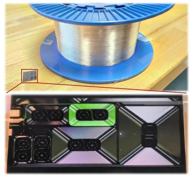
Toggle light / dark theme

Get the latest international news and world events from around the world.

Log in for authorized contributors

Immunotherapy could prevent the loss of neurons in Parkinson’s disease

Parkinson’s disease is characterized by the progressive loss of dopaminergic neurons in a specific brain region known as the substantia nigra. This neuronal degeneration is closely linked to inflammatory processes mediated by microglia, the immune cells of the central nervous system. However, the precise mechanisms that trigger this destructive process are still not fully understood.

Now, in an article published in npj Parkinson’s Disease, researchers from the Institut de Neurociències of the Universitat Autònoma de Barcelona (INc-UAB) and the UAB Department of Biochemistry and Molecular Biology report that brain tissue from Parkinson’s disease patients contains a higher proportion of reactive microglia, meaning cells that are primed to respond. But most importantly, these reactive microglial cells also show an increased density of receptors known as Fc gamma on their membranes.

Study maps 30 rheumatoid arthritis biopsies, linking joint scarring to treatment resistance

Rheumatoid arthritis (RA) is a common autoimmune disease where the body’s immune system mistakenly attacks the lining of its own joints, causing chronic pain, swelling, and stiffness. While there have been remarkable advancements in the treatment of RA with an array of therapies that target inflammation, a large subset of patients (approximately 6–28%) continue to experience difficult-to-manage symptoms of disease even after receiving multiple lines of treatment.

There is a critical need to identify new therapeutic approaches for patients who are refractory to existing treatment options.

By looking closely at the biology of joint tissue, researchers at the Mass General Brigham Department of Medicine conducted a study, published in Nature Immunology, focusing on discovering why some people with rheumatoid arthritis don’t respond well to standard treatments. The paper is titled “Spatial patterning of fibroblast TGFβ signaling underlies treatment resistance in rheumatoid arthritis.”

Scientists Uncover a Hidden Early Stage of Alzheimer’s That They Can Stop

The researchers suggested that higher concentrations of charged ions weaken the interaction between tau proteins and heparin, making cluster formation more difficult. This occurs because charged molecules such as tau and heparin become less able to interact due to electrostatic “screening,” which effectively masks their charges from one another.

A New Direction for Treating Neurodegenerative Disease

These results point toward a different strategy for developing therapies. Rather than attempting to break apart fully formed tau fibrils, future treatments could focus on blocking the reversible precursor stage before irreversible damage takes place. This approach could have implications beyond Alzheimer’s disease, potentially influencing research into other neurodegenerative disorders, including Parkinson’s disease.

Ferroptosis as a therapeutic target in glioblastoma: Mechanisms and emerging strategies

Ferroptosis: a promising therapeutic strategy in glioblastoma👇

✅Glioblastoma multiforme (GBM) is an aggressive brain tumor characterized by rapid growth and resistance to conventional therapies. Recent research highlights ferroptosis, a regulated form of cell death driven by iron-dependent lipid peroxidation, as a novel and promising approach for GBM treatment.

✅One key mechanism underlying ferroptosis in GBM is glutathione depletion. Inhibition of the cystine/glutamate antiporter system (xCT) limits cystine uptake, leading to reduced glutathione synthesis. As a consequence, the antioxidant enzyme GPX4 becomes inactivated, impairing the cell’s ability to detoxify lipid peroxides.

✅Lipid peroxidation is a central event in ferroptosis. Polyunsaturated fatty acids (PUFAs) incorporated into membrane phospholipids are highly susceptible to oxidative damage. Their conversion into peroxidized phospholipids (PL-PUFA-PE) disrupts membrane integrity and drives lethal oxidative stress.

✅Iron metabolism further amplifies ferroptotic signaling in GBM cells. Elevated intracellular iron, particularly the Fe²⁺ pool, catalyzes redox reactions that generate reactive oxygen species (ROS). This iron-driven ROS production accelerates lipid peroxidation and pushes tumor cells toward ferroptotic death.

✅Collectively, glutathione depletion, GPX4 inactivation, uncontrolled lipid peroxidation, and dysregulated iron metabolism converge to induce ferroptosis. Targeting these interconnected pathways offers a potential strategy to overcome therapy resistance and selectively eliminate GBM cells.

A glycolytic shunt via the pentose phosphate pathway is a metabolic checkpoint for nervous system sensory homeostasis and axonal regeneration

Pentose phosphate pathway in axonal regeneration.

Various signaling pathways play an important role in neuronal homeostasis and regeneration.

The researchers in this study determine that the pentose phosphate pathway (PPP) plays a dual role in enabling homeostatic and regenerative adaptations to environmental stimuli and injuries.

They show that sciatic nerve axoplasms are enriched PPP and maintains redox balance via NADPH production but following sciatic nerve injury, the PPP is required for regeneration by fueling ribonucleotide synthesis through ribose-5-phosphate.

However, after spinal cord injury (SCI), PPP remain inactive and neuronal transketolase overexpression or oral ribose supplementation, promotes metabolic reprogramming, restores sensory and motor axonal growth. sciencenewshighlights ScienceMission https://sciencemission.com/pentose-phosphate-pathway-is-a-metabolic-checkpoint


The pentose phosphate pathway plays a dual role in enabling homeostatic and regenerative adaptations to environmental stimuli and injuries and can be leveraged to promote regeneration and recovery after spinal cord injury.

The contribution of the membrane-bound complement regulatory proteins CD46 and CD55 in phases of acute lymphocytic leukemia and acute myelogenous leukemia

As for decay accelerating factor (DAF); also known as CD55, it is a type I cell surface protein that forms a single chain anchored to the membrane by glycosylphosphatidylinositol (GPI). It binds C3b and C4b inhibiting thereby the formation of C3 convertase and decreasing its half-life, thus providing a protective barrier threshold for plasma membranes of normal autologous cells against complement deposition and activation9,10.

The role of the complement system in cancer is complicated and has been debated for long. Malignant transformation is generally accompanied by genetic and epigenetic modifications which drastically alter patterns of glycosylation, cell-surface proteins and phospholipids11. These alterations can be identified by innate and adaptive immune mechanisms that guard the host against cancer development12. This is the known basis of the immune surveillance hypothesis. There is no direct evidence to support the argument that complement is able to eradicate emerging tumors. Nevertheless, taking into consideration that complement is intended for the recognition of non-self-elements, it is assumed that alterations in the tumor cell membranes’ composition render these cells as targets for complement recognition13. However, the relationship between inflammation and cancer is complicated and subject to contradictory forces14. Therefore, while acute responses are considered a vital part of the defense against cancerous cells, continuous inflammation in the tumor microenvironment increases the threat of neoplastic transformation and has several tumor-promoting effects15.

The current study aims at investigating the expression levels of mCRPs; CD46 and CD55 in the acute lymphocytic leukemia and acute myelogenous leukemia and to further elucidate its role in Egyptian cancer patients. To the best of our knowledge this study is one of very few studies tackling the complicated role of the complement system in acute leukemia.

/* */