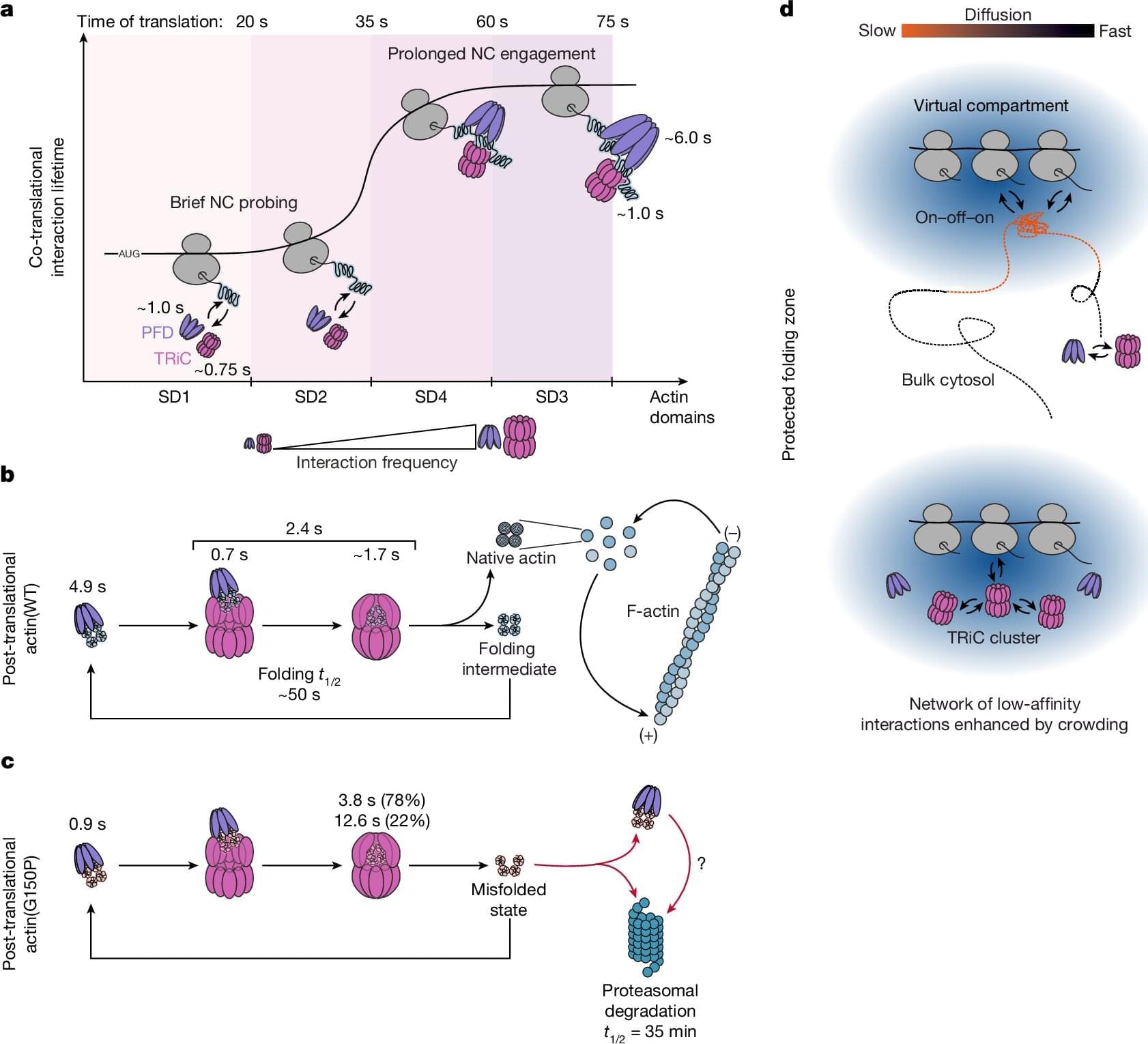
Toggle light / dark theme

Get the latest international news and world events from around the world.

Log in for authorized contributors

Neuron position found less crucial for brain connectivity than once thought

The human brain contains billions of connected neurons that collectively support different mental functions, including the processing of sensory information, the encoding of memories, attention processes, and decision-making. For a long time, neuroscientists have assumed the position of specific neurons in the brain plays a key role in the brain’s connectivity and proper functioning.

Researchers at University of Geneva, INSERM, Ecole Polytechnique Fédérale de Lausanne and other institutes recently gathered evidence that contradicts this long-standing assumption, showing misplaced neurons can still retain their “identity,” connect with other neurons and support the processing of sensory information.

Their paper, published in Nature Neuroscience, could reshape the present understanding of developmental disorders and other conditions linked to the rearrangement of neurons or cortical malformations.

Different Autism Mutations Can Lead to Similar Brain Changes

The shared pathways were linked to neuron maturation, synapse formation, and the control of gene activity. Further analysis pointed to a group of genes involved in organizing DNA and regulating which genes are switched on or off. These genes sit high in the regulatory chain, influencing many downstream processes previously linked to autism.

To test whether this network played an active role, the team reduced the activity of several key regulators using CRISPR-based methods in neural cells. This led to downstream changes similar to those seen in the autism models.

However, organoids from individuals with idiopathic autism showed less consistent changes, likely reflecting the complex and distributed genetic risk seen in most autism cases.

CLN3 mediates chloride efflux from lysosomes

Lysosomes degrade damaged organelles and macromolecules to recycle nutrient components. Lysosomal storage diseases (LSDs) are linked to mutations of genes encoding lysosomal proteins and may lead to age-related disorders, including neurodegenerative diseases. But, how lysosomal dysfunction contributes to neurodegenerative diseases is not clear yet…

The researchers identify CLN3 (ceroid lipofuscinosis, neuronal 3), linked to Batten disease as a conserved lysosomal protein that regulates lysosomal chloride homeostasis, pH, and protein degradation.

Curcumin analog C1 is a natural compound with anti-inflammatory properties could enhance CLN3 activity and improve lysosomal function by activating TFEB. sciencenewshighlights ScienceMission https://sciencemission.com/CLN3-n-chloride-efflux-n-lysosomes


Wang et al. identify CLN3 as a conserved lysosomal protein that regulates lysosomal chloride homeostasis, pH, and protein degradation. Transcription factor EB (TFEB) activation enhances CLN3 function, revealing the TFEB-CLN3 signaling axis as a promising therapeutic target for lysosomal storage disorders.

Resolving DNA origami structural integrity and pharmacokinetics in vivo

Impressive leap forwards for DNA origami: an elegant staple strand proximity ligation method for tracking DNA origami pharmacokinetics in vivo! This approach even allows analysis of stability of subregions within a DNA origami nanostructure. I think DNA origami has a lot of therapeutic potential, so it is exciting to see this solution to one of its translational barriers. Link: https://www.nature.com/articles/s41565-025-02091-z Paper title: “Resolving DNA origami structural integrity and pharmacokinetics in vivo”


Using origami samples in test tubes, we sequentially performed ligation, PCR and polyacrylamide gel electrophoresis (PAGE; Fig. 2a). For both Wrod and Lrod, amplification bands appeared only after ligation (Supplementary Fig. 3) and matched the sizes of single-LSP controls (Fig. 2d, g). When the origami was heat denatured before ligation, no LSP bands were detected (Fig. 2e, h), confirming that proximity ligation requires intact structures. By contrast, we showed that scaffold-targeted qPCR or origamiFISH assays37,38 still detected DNA regardless of the structural state (Fig. 2e, h), emphasizing their inability to distinguish intact origami from degraded origami.

Previous studies have shown that the coating of DNA nanostructures with the oligolysine-PEG polymer can protect them against nucleases and denaturation in low-salt environments, potentially increasing their stability in vivo23. Since PEGylation confers a physical barrier for the interaction of enzymes with DNA helices, we hypothesized that the ligase might also have decreased accessibility to PEGylated origamis. However, our in vitro experiments with PEGylated PEG-Lrod showed comparable ligation and amplification efficiencies to the bare Lrod (Supplementary Fig. 4). Another approach to enhance lattice-based origami stability in low-salt buffers and improved resistance to nucleases is sequence-specific covalent UV crosslinking26. We tested the application of the PLASTIQ protocol to a crosslinked version of the Lrod (UV-Lrod) with the same LSPs as Lrod. We observed a similar amplification pattern when compared to the non-crosslinked Lrod after PAGE electrophoresis of the pooled PCR-amplified LSPs (Extended Data Fig. 1).

Together, these results demonstrate that PLASTIQ reliably detects DNA origami integrity at the single-helix level for both wireframe and lattice designs, and that it is compatible with PEGylated or UV-crosslinked nanostructures.

Advancing regulatory variant effect prediction with AlphaGenome

What makes it special is its versatility. Where older models might only predict how a mutation affects gene activity, AlphaGenome forecasts thousands of biological outcomes simultaneously—whether a variant will alter how DNA folds, change how proteins dock onto genes, disrupt the splicing machinery that edits genetic messages, or modify histone “spools” that package DNA. It’s essentially a universal translator for genetic regulatory language.


AlphaGenome is a deep learning model designed to learn the sequence basis of diverse molecular phenotypes from human and mouse DNA (Fig. 1a). It simultaneously predicts 5,930 human or 1,128 mouse genome tracks across 11 modalities covering gene expression (RNA-seq, CAGE and PRO-cap), detailed splicing patterns (splice sites, splice site usage and splice junctions), chromatin state (DNase, ATAC-seq, histone modifications and transcription factor binding) and chromatin contact maps. These span a variety of biological contexts, such as different tissue types, cell types and cell lines (see Supplementary Table 1 for the summary and Supplementary Table 2 for the complete metadata). These predictions are made on the basis of 1-Mb of DNA sequence, a context length designed to encompass a substantial portion of the relevant distal regulatory landscape. For instance, 99% (465 of 471) of validated enhancer–gene pairs fall within 1 Mb (ref. 12).

AlphaGenome uses a U-Net-inspired2,13 backbone architecture (Fig. 1a and Extended Data Fig. 1a) to efficiently process input sequences into two types of sequence representations: one-dimensional embeddings (at 1-bp and 128-bp resolutions), which correspond to representations of the linear genome, and two-dimensional embeddings (2,048-bp resolution), which correspond to representations of spatial interactions between genomic segments. The one-dimensional embeddings serve as the basis for genomic track predictions, whereas the two-dimensional embeddings are the basis for predicting pairwise interactions (contact maps). Within the architecture, convolutional layers model local sequence patterns necessary for fine-grained predictions, whereas transformer blocks model coarser but longer-range dependencies in the sequence, such as enhancer–promoter interactions.

/* */