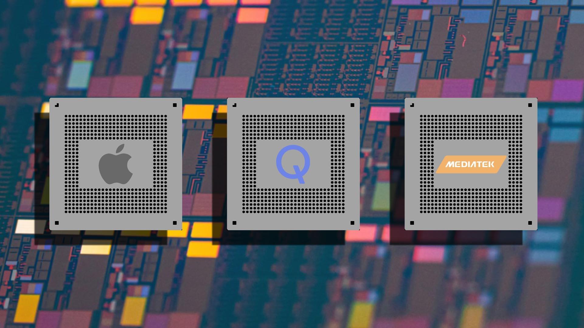
Toggle light / dark theme

Cells are constantly subjected to DNA damage from a range of internal and environmental sources. It is estimated that cells can experience as many as 100,000 DNA lesions per day. One of the most deleterious types of DNA lesions is the DNA double-strand break (DSB). Just one unrepaired DNA DSB may be enough to cause mutations or cell death leading to a wide range of pathologies including cancer, immune deficiency, premature aging and neurodegeneration.

To respond to the array of DNA lesions that occur, cells have developed a complex and coordinated series of steps involving DNA damage recognition, cell cycle arrest and signaling-induced activation of the DNA repair machinery—processes collectively referred to as the DNA damage response (DDR). In recent years, progress has been made in understanding how this process is initiated. However, the later stages of this process, including long range DNA end-resection, are not well understood.

In a new study published in Nature Cell Biology, researchers from Boston University Chobanian & Avedisian School of Medicine, Massachusetts General Hospital (MGH) and Harvard Medical School, identified several uncharacterized chromatin factors (proteins that regulate ) that are recruited to sites of DNA damage, including the gene ZNF280A. Importantly, this gene is hemizygously deleted—meaning one of the two copies of alleles is missing—in a subset of patients with a human developmental syndrome called 22q11.2 distal deletion syndrome.

As the opioid epidemic persists across the United States, a team of researchers from Brown University has developed new diagnostic techniques for detecting opioid compounds in adults with opioid use disorder and infants with neonatal abstinence syndrome.

The new techniques, described in two recently published research studies, could equip with powerful new tools for more effectively treating conditions related to opioid exposure, the researchers say.

In a study published in Scientific Reports, the researchers describe a method that can rapidly detect six different opioid compounds from a tiny amount of serum—no more than a finger prick.

A major medical breakthrough is giving new hope to those with Parkinson’s disease. Stem cell transplants into the brain are showing impressive results, leading to long-term improvements in motor symptoms for the first patients treated… Read more

Our brains begin to create internal representations of the world around us from the first moment we open our eyes. We perceptually assemble components of scenes into recognizable objects thanks to neurons in the visual cortex.

This process occurs along the ventral visual cortical pathway, which extends from the primary at the back of the brain to the temporal lobes.

It’s long been thought that specific neurons along this pathway handle specific types of information depending on where they are located, and that the dominant flow of visual information is feedforward, up a hierarchy of visual cortical areas. Although the reverse direction of cortical connections, often referred to as feedback, has long been known to exist, its functional role has been little understood.

People who have damage to a specific part of their brains are more likely to be impulsive, and new research has found that damage also makes them more likely to be influenced by other people.

In a new study published in PLOS Biology, a research team found that damage to distinct parts of the (mPFC) was linked to being influenced by impulsive decision-making by others, while another region was causally linked with choosing a smaller reward earlier rather than waiting for a larger prize.

The team from the University of Birmingham, University of Oxford and Julius-Maximilians-University Würzburg worked with participants with brain damage to assess whether they were more likely to be influenced by other people’s preferences.