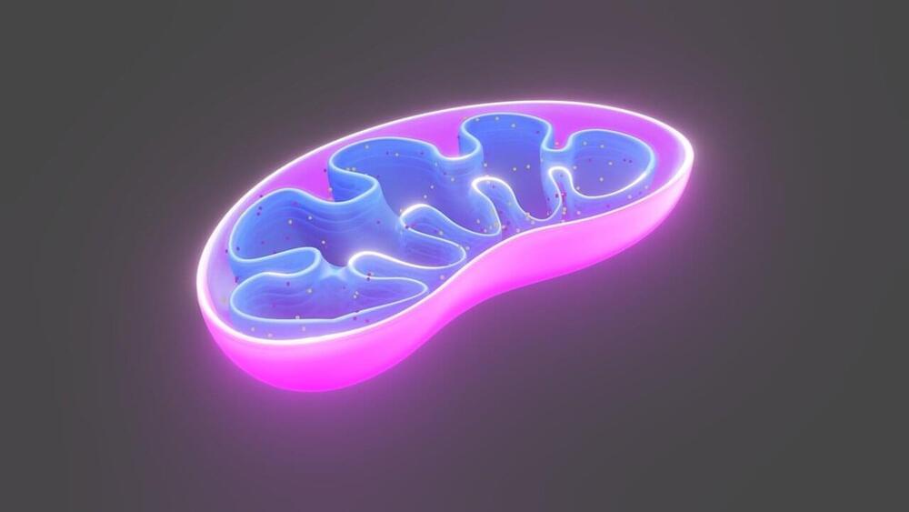
Toggle light / dark theme

Growing organs in the lab is an enduring sci-fi trope, but as stem cell technology brings it ever closer to reality, scientists are beginning to contemplate the ethics governing disembodied human tissue.

So-called organoids have now been created from gut, kidney, pancreas, liver and even brain tissue. Growing these mini-organs has been made possible by advances in stem cell technology and the development of 3D support matrices that allow cells to develop just like they would in vivo.

Unlike simple tissue cultures, they exhibit important structural and functional properties of organs, and many believe they could dramatically accelerate research into human development and disease.

Read more

Could we eventually see a day where we have cell circuitry nanobot pill that eliminates hunger and obesity as replacement to gastric bypasses? Maybe.


The human body responds to starving conditions, such as famine, to promote the chance of survival. It reduces energy expenditure by stopping heat production and promotes feeding behavior. These “hunger responses” are activated by the feeling of hunger in the stomach and are controlled by neuropeptide Y (NPY) signals released by neurons in the hypothalamus. However, how NPY signaling in the hypothalamus elicits the hunger responses has remained unknown.

Sympathetic motor neurons in the medulla oblongata are responsible for heat production by brown adipose tissue (BAT). Researchers centered at Nagoya University have now tested whether the heat-producing neurons respond to the same hypothalamic NPY signals that control hunger responses. They injected NPY into the hypothalamus of rats and tested the effect on heat production. Under normal conditions, blocking inhibitory GABAergic receptors or stimulating excitatory glutamatergic receptors in the sympathetic motor neurons induced heat production in BAT. After NPY injection, stimulating glutamatergic receptors did not produce heat, but inhibiting GABAergic receptors did. The study was recently reported in Cell Metabolism.

Retrograde and anterograde tracing with fluorescent dyes revealed which brain region provided the inhibitory GABAergic inputs to heat-producing motor neurons.

The brain is the fattiest organ in your body made up of 60% fat, the dry part that is. 75% of your brain is actually water which houses 100,000 miles of blood vessels that use up 20% of all your oxygen and blood. It’s an amazing piece of hardware. Of all the moonshot projects out there, the ones that relate to augmenting the brain are perhaps the most fascinating. Companies like Kernel have actually succeeded in writing long-term memories to a chip – well, at least 80% of them. When that number hits 100%, the sky is the limit to what we can do with the brain.

If you want a graphic image of what the future holds, imagine a robotic arm on top of your table (no wires) moving its fingers or trying to grab something powered only by someone’s thought. After all those Terminator movies, this could be a bit creepy. You may not get Terminator at your doorstep just yet, but someone with neuroprosthesis might just be ringing your doorbell a few years from now.

Neuroprosthetics or neuroprosthesis is a field of biomedical engineering and neuroscience concerned with the development of neural prostheses which are a series of devices that can substitute your brain’s motor, sensory or cognitive functionality that might have been damaged as a result of an injury or a disease.

Read more

In Brief:

  • The Multiple Sclerosis Foundation estimates that more than 400,000 people in the United States and about 2.5 million people around the world have MS.
  • A new drug, Ocrelizumab, is the first known drug shown to work against the primary progressive form of MS by altering the immune system to slow damage to the brain.

Multiple sclerosis (MS) is an unpredictable and potentially disabling disease that cripples the central nervous system. It’s a widespread neurological condition that hits young adults, usually between the ages of 20 and 40, caused by an immune system disorder that mistakes a part of the brain as a hostile foreign object and attacks it. Though there are treatments available, particularly for its second state, multiple sclerosis remains incurable.

Read more

Neuroscientists at the University of Bristol are a step closer to understanding how the connections in our brain which control our episodic memory work in sync to make some memories stronger than others. The findings, published in Nature Neuroscience, reveal a previously unsuspected division of memory function in the pathways between two areas of the brain, and suggest that certain subnetworks within the brain work separately, to enhance the distinctiveness of memories.

The team studied the hippocampus and prefrontal cortex—two regions of the brain critical to function—as damage in these areas can induce severe memory loss.

Both areas are connected by a complex network of direct and indirect pathways, and the challenge has been until now, how to identify the precise routes through which these brain regions interact in memory formation.

Read more

In Brief

  • By mimicking the way neurons fire in the hippocampus during natural memory creation, a brain implant was used to successfully plant memories in the brains of rats.
  • Though human implementation is far off, this breakthrough in cracking the hippocampus’ mathematical “memory code” has very important implications for health and research.

Memories are the faintest, most ethereal wisps of our neurophysiology — somehow, the firing of delicate synapses and the activation of neurons combine to produce the things we remember. The sum of our memories make us who we are; they are us, in every way, and without them we cease to be.

So it’s needless to say that there’s a pretty significant premium on discovering new ways to combat memory loss. Most of these involve physiological and biological methods, but some scientists, such as Theodore Berger of the University of Southern California, are beginning to turn toward technology. If any of these methods are successful, it would mean the possibility of perfect lifelong memory recall.

Read more

January 20, 2017 — Researchers have developed a high-resolution, interactive anatomic brain-mapping atlas they say can overcome the limitations of functional MRI (fMRI) and expand the modality’s value to conventional MR brain imaging and standard MRI applications.

The Gibby-Cvetko atlas is designed to segment the brain into finite anatomic regions with a resolution of 1 mm or less to correct for variations in the brain sizes of patients and better delineate the location of cortical structures and skull morphology.

“Having a high-resolution, interactive, quantitative brain atlas that we warp to fit the patient and run inside a PACS improves accuracy and speed of reading fMRI studies,” said co-developer Dr. Wendell Gibby, an adjunct professor of radiology at the University of California, San Diego. “It is a big step toward routine utilization of fMRI in clinical practice.”

Read more