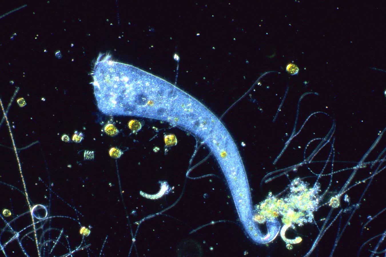
A trumpet-shaped, single-celled organism seems able to predict one thing will follow another, hinting that such associative learning emerged long before multicellular nervous systems
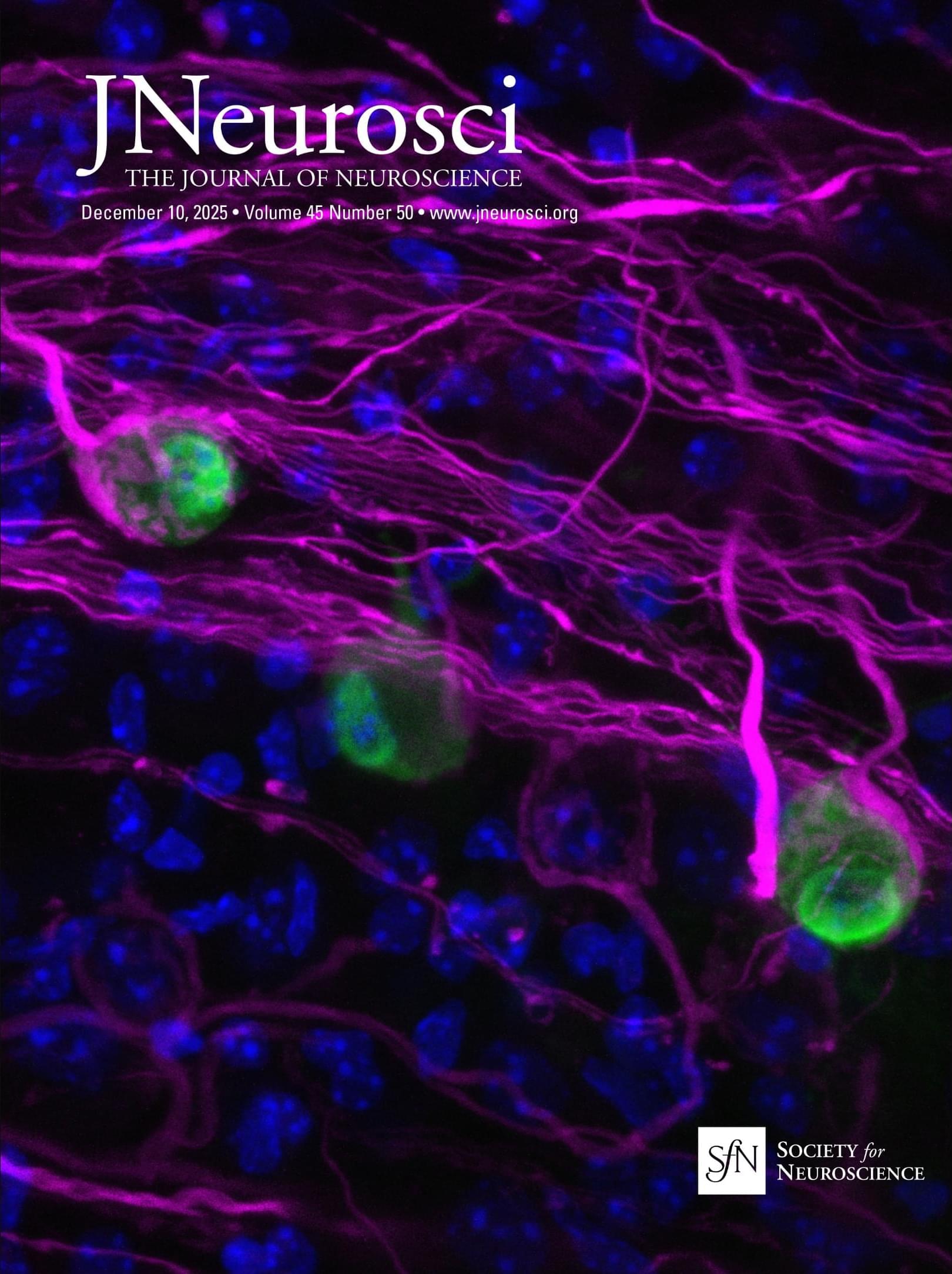
JNeurosci: Martin-Correa et al. combined high-end volumetric electron microscopy and axon labeling methods to measure the synapses established in adult mouse somatosensory area 2 by specific thalamic cell populations.
▶️
The synaptic circuits established by thalamocortical axons from the ventral posteromedial (VPM) and posterior (Po) nuclei in the first somatosensory cortex have been mapped in high detail as they are a prime model in functional and modeling studies of the interactions between the thalamus and cerebral cortex. In addition, VPM and Po neurons innervate the second somatosensory area (S2), but the synaptic organization of their axons in this area remained essentially unknown. On adult male mice, we combined axon labeling with serial section transmission electron microscopy and focused ion beam-scanning electron microscopy to measure and compare functionally relevant structural parameters of synaptic boutons (SBs), e.g., bouton and mitochondrial volume, vesicle pool size, as well as postsynaptic density (PSD) distribution and size.
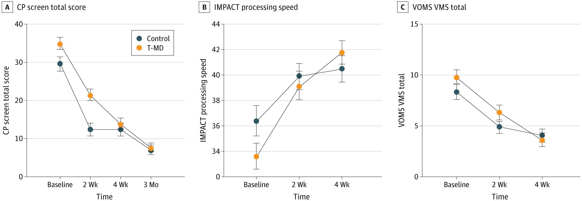
RCT: Among adults with mTBI, targeted multidomain interventions and usual care behavioral management resulted in similar improvements in overall symptom severity and patient-perceived recovery over 4 weeks.
Question Is a targeted multidomain (T-MD) intervention more effective than a standardized behavioral management intervention in adults with mild traumatic brain injury (mTBI)?
Findings In this randomized clinical trial of 162 patients, both the T-MD and behavioral management groups experienced similar improvements in mTBI symptom severity and perceived improvement in primary outcomes over 4 weeks.
Meaning The findings suggest that both T-MD and behavioral management are effective for improving global mTBI symptoms.

Scientists have plenty of ideas about why aging impairs memory. Reductions in blood flow in the brain, shrinking brain volume, and malfunctioning neural repair systems have all been blamed. Now, new research in mice points to another possible culprit: microbes in the gut.
In a new study, scientists show how a bacterium that is particularly common in older animals can drive memory loss. This microbe makes compounds that impair signaling along neurons connecting the gut with the brain, dampening activity in brain regions associated with learning and memory, the team found.
Research suggests the microbiome may contribute to cognitive decline—but its relevance in humans is unclear.


New in JNeurosci: fMRI study from Kim et al. reveals that babies with congenital heart disease have altered sensorimotor and limbic brain networks that cardiovascular surgery improves.
▶️
Congenital heart disease (CHD) affects approximately 1% of live births in the United States and is the most prevalent congenital disorder. Despite advances in neonatal cardiovascular surgery improving survival, neurodevelopmental impairments remain prevalent, impacting motor skills, social behavior, and executive function. Motor deficits and long-term challenges in emotional regulation and memory are particularly common. Recent research using resting-state functional MRI (rs-fMRI) has revealed disorganized brain networks in newborns with CHD. However, those few prior rs-fMRI studies examining the impact of CHD have relied on predefined brain parcellations to compare group-level connectivity, limiting the ability to capture spatial alterations in neonatal brain networks in CHD. Understanding these network-level changes is critical for elucidating mechanisms of neurodevelopmental impairment and identifying early biomarkers of risk. To address these gaps, our study introduces two conceptual advances: 1) a data-driven approach to investigate atypical brain network development in high-risk CHD and 2) the use of a population-sized, independent dataset of healthy newborns to derive a normative set of neonatal brain networks. By analyzing a large rs-fMRI of human newborns (N=448; 219 females and 229 males), we identify atypical brain activity in the sensorimotor and limbic networks of newborns with complex CHD. Notably, before cardiovascular surgery, these networks are split into left and right hemispheric subnetworks. Postoperatively, these components coalesce into a singular, symmetric pattern resembling networks observed in healthy neonates. Our study highlights the potential of rs-fMRI to detect subtle, early functional disruptions in CHD and may inform future biomarkers of neurodevelopmental risk.
Significant Statement Congenital heart disease, the most common congenital disorder, affects 1% of live births and is associated with persistent neurodevelopmental impairments despite improved surgical survival. These deficits, including motor, socio-emotional, and cognitive challenges, may stem from early brain network disruptions. Prior resting-state fMRI studies in CHD relied on predefined parcellations, limiting detection of subtle spatial alterations. In this study, we used a data-driven approach and leveraged an independent normative dataset to define resting-state networks. Comparing CHD patients and healthy controls against these independently derived networks, we reveal atypical sensorimotor and limbic network organization preoperatively, which normalizes post-surgery. These findings highlight the potential of rs-fMRI to identify early biomarkers of neurodevelopmental risk and guide targeted interventions in this high-risk population.
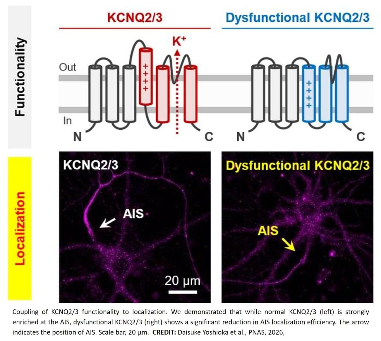
In a study published recently in PNAS, researchers have revealed the relationship between KCNQ2/3 channel functionality (i.e., how well they work to control electrical signals in neurons) and localization (i.e., where they are found inside a cell), with important implications for the treatment of these epileptic disorders.
For KCNQ2/3 channels to work properly in the brain, they must have full functionality and be located in the correct cellular region – specifically the axon initial segment (AIS), the site in neurons where electric signals are first triggered, controlling nerve cell activity. This led the research team to wonder: does the functionality of KCNQ2/3 channels affect their cellular localization, or are the two not linked at all?
To investigate this potential association, the research team first genetically engineered the functionality of the channels, and then used channel trafficking imaging to visualize whether the channels were taken to their location in the AIS. In this way, they demonstrated that KCNQ2/3 functionality was indeed linked to its trafficking to the correct cellular localization. What’s more, when they used single-molecule imaging, they could see that reduced KCNQ3 functionality actually reduced the AIS localization of KCNQ2/3 by altering the entire trafficking pathway.
“Because we already knew that the localization of KCNQ2/3 to the AIS is regulated by a protein known as ankyrinG, or ankG, we next decided to explore the interactions between full-length KCNQ3 and ankG,” explains lead author of the study. “We found that the active conformation of KCNQ3 was essential for its stable binding to ankG, further confirming that functional KCNQ2/3 is needed to ensure its proper accumulation at the AIS.”
Together, these findings highlight the mechanisms underlying the important link between KCNQ2/3 functionality and localization, and provide clues about how their alterations might affect neuronal excitability. ScienceMission sciencenewshighlights.
Potassium KCNQ2/3 channels are crucial for suppressing the excitability of brain cells, or neurons. When these channels don’t work properly, they can cause specific types of epilepsy like benign familial neonatal convulsions and early infantile epileptic encephalopathy.
Reprogramming in melanocortin modulates diet-induced obesity.
Hypothalamic proopiomelanocortin (POMC) neurons promote satiety, while agouti-related peptide (AgRP) neurons drive hunger and maintain energy balance.
However, it is not clear how the system is diversified developmentally.
The researchers in this study show that transcription factor Otp act as a developmental ‘‘switch’’ in the hypothalamus and determines whether immature neurons become appetite suppressing (POMC) or appetite stimulating (AgRP).
Disrupting this switch reshapes feeding behavior and protects mice from obesity, revealing how early life programming shapes lifelong metabolic health. sciencenewshighlights ScienceMission https://sciencemission.com/melanocortin-neurons-modulates-diet-induced-obesity
Xu et al. show that a developmental “switch” in the hypothalamus determines whether immature neurons become appetite suppressing or appetite stimulating. Disrupting this switch reshapes feeding behavior and protects mice from obesity, revealing how early-life programming shapes lifelong metabolic health.