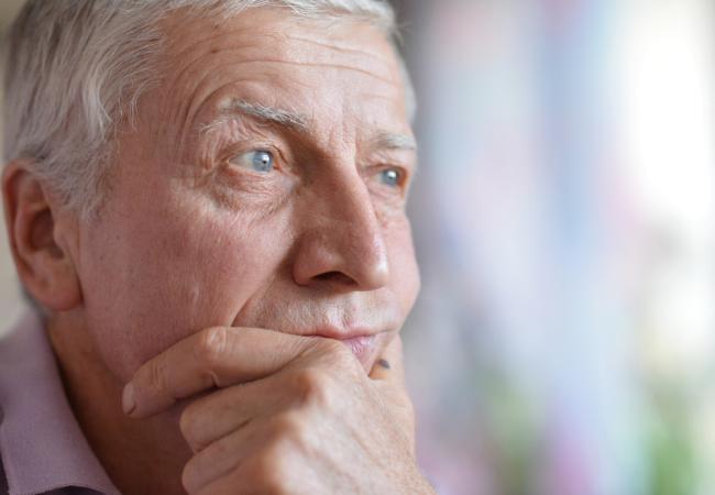
A newly identified enzyme target may open an alternative path for treating Alzheimer’s disease.
Biohacks and brain implants are coming fast. From memory upgrades to neural meshes, how will implant culture reshape identity, economics, and civilization itself?
Get Nebula using my link for 50% off an annual subscription: https://go.nebula.tv/isaacarthur.
Watch my exclusive video Lazarus Protocols: https://nebula.tv/videos/isaacarthur–… out Tech Alter: https://nebula.tv/techaltar?ref=isaac… and Day Pass: https://nebula.tv/daypass?ref=isaacar… 🛒 SFIA Merchandise: https://isaac-arthur-shop.fourthwall… 🌐 Visit our Website: http://www.isaacarthur.net ❤️ Support us on Patreon: / isaacarthur ⭐ Support us on Subscribestar: https://www.subscribestar.com/isaac-a… 👥 Facebook Group:
/ 1,583,992,725,237,264 📣 Reddit Community:
/ isaacarthur 🐦 Follow on Twitter / X:
/ isaac_a_arthur 💬 SFIA Discord Server:
/ discord Credits: Biohacks & Brain Mods — The Coming Age of Implant Culture Written, Produced & Narrated by: Isaac Arthur Edited by: Chris Gray & Donagh Broderick Select imagery/video supplied by Getty Images Chapters 0:00 Intro 1:28 The First Layer – The Implants Already Here 3:08 The Early Adoption Phase – Niche Enhancements 5:27 Safety vs Freedom 8:25 Implant Economics 14:31 The Social Shift – The Rise of Implant Culture 20:04 Minds in Motion – Cognitive Architecture as a Design Space 21:50 Implant Maximalism – Full-Stack Upgrades 23:03 Nebula 24:09 Post-Implant Society – Upgrades as Life Stages 25:40 The Far Horizon – When Implants Become Civilization.
Check out Tech Alter: https://nebula.tv/techaltar?ref=isaac…
and Day Pass: https://nebula.tv/daypass?ref=isaacar…
🛒 SFIA Merchandise: https://isaac-arthur-shop.fourthwall…
🌐 Visit our Website: http://www.isaacarthur.net.
❤️ Support us on Patreon: / isaacarthur.
⭐ Support us on Subscribestar: https://www.subscribestar.com/isaac-a…
👥 Facebook Group: / 1583992725237264
📣 Reddit Community: / isaacarthur.
🐦 Follow on Twitter / X: / isaac_a_arthur.
💬 SFIA Discord Server: / discord.
Credits:
Biohacks & Brain Mods — The Coming Age of Implant Culture.
Written, Produced & Narrated by: Isaac Arthur.
Edited by: Chris Gray & Donagh Broderick.
Select imagery/video supplied by Getty Images.
Chapters.
0:00 Intro.
1:28 The First Layer – The Implants Already Here.
3:08 The Early Adoption Phase – Niche Enhancements.
5:27 Safety vs Freedom.
8:25 Implant Economics.
14:31 The Social Shift – The Rise of Implant Culture.
20:04 Minds in Motion – Cognitive Architecture as a Design Space.
21:50 Implant Maximalism – Full-Stack Upgrades.
23:03 Nebula.
24:09 Post-Implant Society – Upgrades as Life Stages.
25:40 The Far Horizon – When Implants Become Civilization.
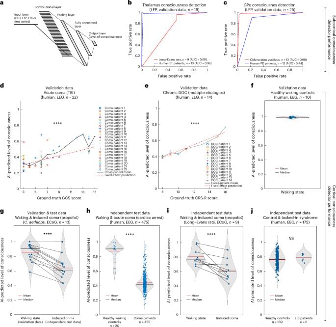
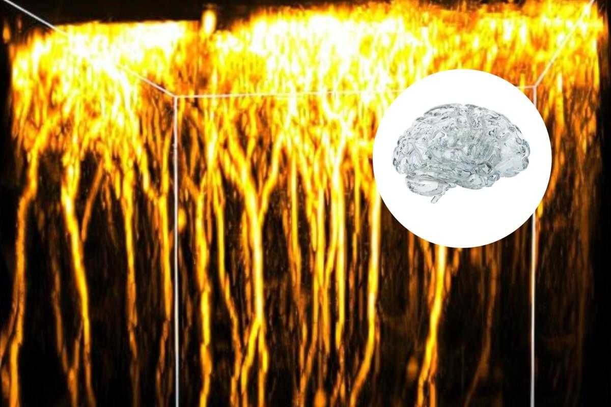
Researchers led by UCLA have developed an adversarial AI framework that may help explain how consciousness breaks down after brain injury — and how it might one day be restored. Published in Nature Neuroscience, the study used deep neural networks trained on more than 680,000 neuroelectrophysiology samples and validated findings across 565 patients, healthy volunteers, and animals. The model identified specific circuit-level disruptions linked to disorders of consciousness, including the basal ganglia indirect pathway and altered inhibitory cortical wiring.
What makes this so important is that it pushes consciousness research closer to mechanism. Instead of only asking what consciousness is, this kind of work asks: what specific brain circuitry fails when consciousness is lost, and can that failure be targeted? The study also identified high-frequency stimulation of the subthalamic nucleus as a promising intervention, supported by human electrophysiological data. This is the kind of neuroscience that makes consciousness feel less like pure philosophy — and more like something we may eventually model, test, and repair.
Abstract: Nature Neuroscience Adversarial AI reveals mechanisms and treatments for disorders of consciousness.
Toker et al. present an AI framework that identifies mechanisms of consciousness. The model predicts new drivers of unconsciousness and identifies subthalamic nucleus stimulation as a potential therapy for disorders of consciousness.
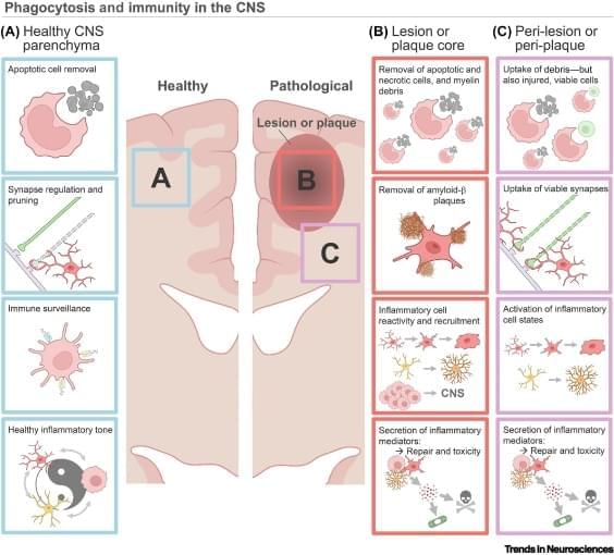
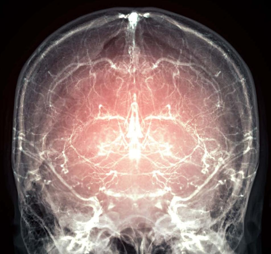
Brain phagocytosis and neuroinflammation.
Phagocytes in the central nervous system (CNS), including astrocytes, microglia, and macrophages, shape development and homeostasis by pruning synapses and removing apoptotic debris.
Phagocytosis is mediated by various ligand–receptor dyads and signaling pathways, enabling CNS phagocytes to respond to neuroimmune shifts across the lifespan and during pathology.
Phagocytosis pathways regulate recovery in various models of CNS pathology, including multiple sclerosis, CNS injury, ischemic stroke, and age-associated neurodegeneration.
Phagocytosis pathways are intimately integrated with the inflammatory cell state and remove viable cells in pathology-adjacent tissue, highlighting the complexity of targeting these systems.
To maximize benefit and minimize off target damage, new phagocytic-based approaches should optimize drug delivery timing and location, tailored to each CNS pathology. sciencenewshighlights ScienceMission https://sciencemission.com/resolution-of-inflammation
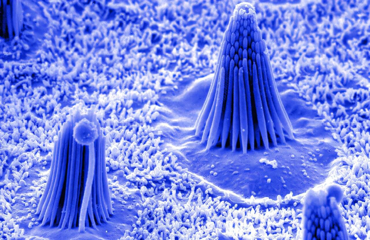
In the intricate machinery of the inner ear, hearing begins with a protein that moves a few billionths of a meter up to 100,000 times per second. That protein, called TMC1, sits at the tips of sensory hair cells deep in the snail-shaped cochlea. When sound waves move these microscopic hairs, TMC1 acts as a channel, opening and allowing charged particles to flow into the cell and trigger an electrical signal to the brain.
Without TMC1, that signal never starts. Mutations in the TMC1 gene are a well-known cause of hereditary hearing loss in humans. Because of this central role, TMC1 is an attractive target for researchers designing gene therapies aimed at restoring hearing. Several groups are testing ways to supply working copies of the gene or fix harmful mutations.
For these efforts to be safe and effective, scientists need to know in detail how TMC1 is built, how it opens, and which parts of the protein are most sensitive to change. However, the hair-cell system that includes TMC1 is so complex, sensitive, and hard to access that it is notoriously difficult to take apart and study directly.
Neurotechnology pioneers @gcourtine and @jocelynebloch are redefining what recovery looks like for people with spinal cord injuries. With a combination of neurosurgery, innovative engineering and AI,…
Year 2025
Cryopreserving the adult brain is challenging due to damage from ice formation, and traditional freezing methods fail to maintain neural architecture and function. Vitrification offers a promising alternative but has not been surveyed in the brain. Here, we demonstrate near-physiological recovery of the adult murine hippocampus after vitrification of brain slices and of the whole brain in situ. Key features of the hippocampus are preserved, including structural integrity, metabolic responsiveness, neuronal excitability, and synaptic transmission and plasticity. Notably, hippocampal long-term potentiation was well preserved, indicating that the cellular machinery of learning and memory remains operational. These findings extend known biophysical limits for cerebral hypothermic shutdown by demonstrating recovery after complete cessation of molecular mobility in the vitreous state. This suggests that the brain can be arrested in time and then reactivated, opening avenues for potential clinical applications.
Significance Statement While the brain is considered exceptionally sensitive, we show that the hippocampus can resume normal electrophysiological activity after being rendered completely immobile in a cryogenic glass. The work extends known biophysical tolerance limits for the brain from the hypothermic to the cryogenic range and establishes a protocol for its long-term storage in a viable state.
The authors have declared no competing interest.
An entire mammalian brain has been successfully preserved using a technique that will now be offered to people who are terminally ill. The intention is to preserve all the neural information thought necessary to one day reconstruct the mind of the person it once belonged to.
“They would need to donate their brain and body for scientific research,” says Borys Wróbel at Nectome in Portland, Oregon, a research company focused on memory preservation. “But what we are offering, as a company, is for their body and brain to be kept, essentially indefinitely, in the hope that sometime, in the future, it would be possible to read out the information from the brain and reconstruct the person… to allow them to continue, in effect, with their life.”
When it comes to preserving the minute architecture of the brain, timing is critical. Within minutes of blood no longer circulating, enzymes break down neurons and cells start digesting themselves.
Image: Samunella/Science Photo Library
A pig’s brain has been frozen with its cellular activity locked in place and minimal damage. Some believe the same could be done with the brains of people with a terminal illness, so their mind can be reconstructed and they can “continue with their life”