New research suggests that tubulin may help prevent the toxic protein clumps associated with Alzheimer’s and Parkinson’s diseases.
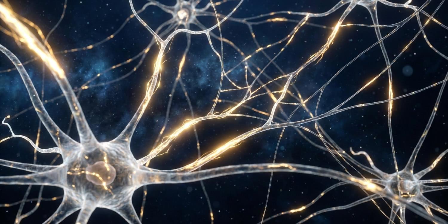
Yet most implants require extensive surgery and risk damaging the brain’s delicate tissue. The new technology would avoid these downsides by building electrodes directly at the target.
“Our work points to a future where doctors could ‘grow’ soft, wire-free electronic interfaces inside the brain using the patient’s own blood, then gently dial brain activity up or down from outside the head using harmless near-infrared light,” study author Krishna Jayant said in a press release.
The brain produces every one of our sensations, movements, emotions, and decisions. Scientists have long sought to decode and manipulate its activity with a range of hardware.
How cancer cells exploit the tumor microenvironment to alleviate oxidative stress remains largely unclear. Zhang et al. find that nociceptive neurons, via secretion of EREG, protect HNSCC against oxidative stress-induced cell death. Targeting nociceptive neurons improves the therapeutic efficacy of chemotherapies, including cisplatin.
The struggle is real when word retrieval goes wrong: see what’s happening in the brain during tip-of-the-tongue moments
The treatment of depression has long faced the challenges of a low treatment rate, significant drug side effects and a high relapse rate. Recent studies have revealed that the gut microbiota and neuronal mitochondrial dysfunction play central roles in the pathogenesis of depression: the gut microbiota influences the course of depression through multiple pathways, including immune regulation, HPA axis modulation and neurotransmitter metabolism. Mitochondrial function serves as a key hub that mediates mood disorders through mechanisms such as defective energy metabolism, impaired neuroplasticity and amplified neuroinflammation. Notably, a bidirectional regulatory network exists between the gut microbiota and mitochondria: the flora metabolite butyrate enhances mitochondrial biosynthesis through activation of the AMPK–PGC1α pathway, whereas reactive oxygen species produced by mitochondria counteract the flora composition by altering the intestinal epithelial microenvironment. In this study, we systematically revealed the potential pathways by which the gut microbiota improves neuronal mitochondrial function by regulating neurotransmitter synthesis, mitochondrial autophagy, and oxidative stress homeostasis and proposed the integration of probiotic supplementation, dietary fiber intervention, and fecal microbial transplantation to remodel the flora–mitochondrial axis, which provides a theoretical basis for the development of novel antidepressant therapies targeting gut–brain interactions.
Depression is a disorder that severely affects the mental health of the global population and is characterized by persistent low mood, loss of interest and cognitive dysfunction (GBD 2017 Disease and Injury Incidence and Prevalence Collaborators, 2018; COVID-19 Mental Disorders Collaborators, 2021; Salari et al., 2020). Globally, depression is one of the leading causes of mental disability. According to the World Health Organization (WHO), the global prevalence of depression is approximately 4.4%, which means that more than 300 million people worldwide suffer from depression (Xu et al., 2024). In addition, depression is one of the major causes of suicide deaths, with nearly 800,000 people worldwide dying by suicide each year (World Health Organization, 2021).
MS is typically diagnosed when clinicians see lesions in the myelin-rich white matter of the brain on MRI scans. White matter is made of the nerves that link brain cells and it looks white on a brain scan.
The brain’s grey matter, which houses the “bodies” of the brain cells, can also have MS lesions, especially in its outer layers. These lesions are less common — and harder to see on a brain scan — but they are a sign of chronic and disabling MS.
The scientists wanted to learn more about the neurons that died in these grey-matter lesions, which express a gene called CUX2. In the first study, they looked at developing mouse brains to see how CUX2 neurons are born. This occurs early in life, when the brain is growing quickly, putting cells under tremendous stress.
The cells relied on a mechanism to repair their DNA as they rapidly multiplied, fanned out into the far reaches of the brain and wired up with one another. The mechanism depends on a stress-response gene called ATF4 to keep chromosomes intact. When the team removed ATF4, the growing neurons were rife with DNA damage, and this prevented the frontal part of the brain from forming.
In the second study, the team found DNA damage in grey matter lesions from people with MS involving the same neurons.
In mouse models of MS, the researchers saw that inflammation sparked chemical reactions that damaged DNA in CUX2 neurons. The repair systems that protect these neurons from the stresses of development could no longer keep up; and this led to brain damage.
Together, the two studies outline the natural way the brain’s outer layer neurons cope with DNA damage — and how that system breaks down in MS. ScienceMission sciencenewshighlights.
Scientists from the Lee Kong Chian School of Medicine at Nanyang Technological University, Singapore, have uncovered how different brain regions work together to enable self-control—the ability to suppress impulsive behaviors and wait for the right moment to act. Their findings advance the understanding of conditions such as attention-deficit hyperactivity disorder (ADHD) and addiction, and could lead to more effective management of these disorders.
According to the researchers, this is the first time that this interplay underlying self-control has been found in the brain. The findings were reported in Science Advances.
A study by investigators at Mass General Brigham has found that a blood test of plasma phosphorylated tau 217 (pTau217), an Alzheimer’s disease biomarker, can predict the progression of amyloid PET scan changes and cognitive decline in cognitively healthy older adults. The findings may help push back the clock to enable simpler, earlier disease prediction and indicate who may be at risk for cognitive decline. The results are published in Nature Communications.
“We used to think that PET scan detection was the earliest sign of Alzheimer’s disease progression, revealing amyloid accumulation in the brain 10 to 20 years before symptoms appear,” said lead author Hyun-Sik Yang, MD, a neurologist with Mass General Brigham Neuroscience Institute and an associate member of the Broad Institute of MIT and Harvard. “But now we are seeing that pTau217 can be detected years earlier, well before clear abnormalities appear on amyloid PET scans.”
Last year, the U.S. Food and Drug Administration cleared the first blood test for Alzheimer’s disease, paving the way for a cheaper, less invasive alternative to lumbar punctures and PET scans. The new study by Yang and colleagues adds important evidence about the predictive potential of these kinds of blood tests.
Background Raised cardiac troponin-I is a common finding in patients hospitalised with acute viral infections, including but not limited to COVID-19. This often occurs in the absence of overt myocardial injury presenting a challenge for interpretation. The mechanisms underlying troponin elevation are uncertain.
Methods The CISCO-19 (Cardiovascular Imaging in SARS-CoV-19) study (NCT04403607) is a prospective, multicentre cohort study, in which hospitalised PCR-confirmed COVID-19 participants (N=267) underwent multisystem evaluation at enrolment and at 28–60 days. The study incorporated plasma proteomics (SOMAscan V.4.1), cardiovascular MRI and clinical biomarkers. Of these, 211 had baseline plasma proteomic data and 185 completed follow-up sampling. Matched proteomic and imaging data were available for 155 participants (mean age: 55 years (SD 12); 43% female).
Results A high likelihood of myocarditis was identified in 13.2% (N=21/159) of participants. High-sensitivity troponin-I was modestly elevated at enrolment (median 3 ng/L; IQR 2–6; n=159). Among males (n=90), 9.3% had a high-sensitivity troponin that exceeded 34 ng/L. Among females (n=69), 4.5% exceeded 16 ng/L. Smooth muscle myosin light chain proteins were downregulated at follow-up (log2 fold change −0.12 to −0.6; all adjusted p0.02) and positively correlated with high-sensitivity troponin-I, but not N-terminal brain natriuretic peptide or cardiac MRI indices (n=155).