Cortical Labs says the stunt points toward a new kind of low-power computing—and perhaps a new way to study neurological drugs


A new study from the University of Geneva points to the brain’s waste-clearance system — the glymphatic system — as a possible piece of the psychosis puzzle. In people with 22q11.2 deletion syndrome, a high-risk genetic condition, researchers found developmental differences in an MRI-derived marker linked to glymphatic function, along with associations to hippocampal excitation/inhibition balance and psychosis vulnerability.
A team from UNIGE shows that early alterations in the brain’s clearance system could contribute to vulnerability to psychosis.
How can we explain the onset of psychotic symptoms characteristic of schizophrenia? Despite their major and often irreversible impact on intellectual abilities and autonomy, the biological mechanisms that precede their emergence remain poorly understood. A team from the Department of Psychiatry at the Faculty of Medicine and the Synapsy Center for Neuroscience Research in Mental Health at the University of Geneva (UNIGE) provides new insight into this question. Early dysfunction of the glymphatic system, the network responsible for removing waste from the brain, could be a key vulnerability factor. This research has been published in Biological Psychiatry: Global Open Science.
Hallucinations and delusions are among the characteristic psychotic symptoms of schizophrenia spectrum disorders, which may also be accompanied by social withdrawal and cognitive decline. These disorders, considered neurodevelopmental conditions, most often emerge during adolescence or early adulthood and have an estimated prevalence of 0.5–3% in the general population.

Liu et al. present via https://bit.ly/4bV6X0s (Original research, Hepatology section).
A major step forward for translational research, this study shows that human organoid systems can support replication of multiple hepatitis E virus genotypes—offering a powerful new platform for studying infection and testing therapies.
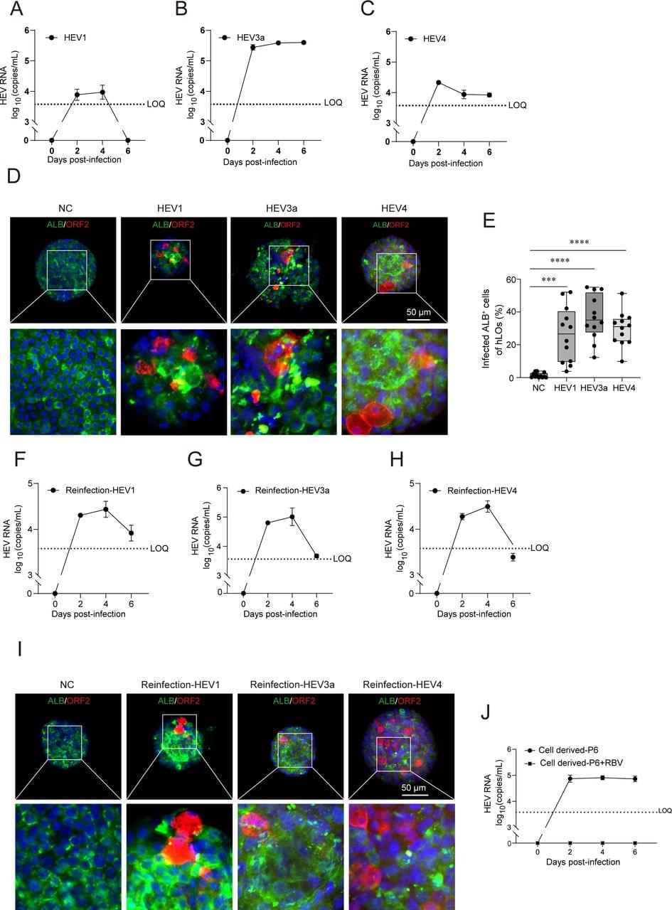
Background Hepatitis E virus (HEV), the leading global cause of acute viral hepatitis, lacks robust in vitro models for virology and pathogenesis research.
Objective We evaluated induced pluripotent stem cell (iPSC)-induced human liver, intestinal and brain organoids (hLOs, hIOs and hBOs) as platforms for HEV infection and replication.
Methods Multilineage organoids were infected with clinical HEV genotypes 1, 3 and 4. Viral tropism, host responses and antiviral efficacy were assessed.
Results All organoids supported the complete life cycle of HEV. hLOs exhibited infection in hepatocytes, cholangiocytes, macrophages and stellate cells, accompanied by elevated interleukin-6 levels, impaired hepatic function (reduced secretion of albumin and Factor IX) and increased levels of alanine aminotransferase and aspartate aminotransferase, indicating hepatocellular injury.
Background Fluid restriction is a commonly prescribed non-pharmacological intervention in the management of heart failure (HF). However, data on its efficacy and safety are scarce. Recent randomised clinical trial (RCT) data prompt reassessment of the available evidence.
Methods CINAHL, EMBASE, PubMed and the Cochrane Library were searched up to 1 May 2025. RCTs were included if adults with HF were randomised to fluid restriction in comparison to a liberal or unrestricted intake, less strict restriction or usual care. Outcomes of interest were mortality, HF hospitalisation, quality of life (QoL), thirst distress, New York Heart Association (NYHA) class and N-terminal pro-Brain Natriuretic Peptide (CRD42022292319). No meta-analysis was performed due to high heterogeneity of the included trials.
Results In total, four RCTs were included, comprising 682 randomised inpatient, recently discharged and stable outpatient patients (ranging from 46 to 504 patients per trial). Only one study had a low risk of bias. None of the four trials found a significant difference in mortality or HF hospitalisations. For QoL, the results are contradictory, but overall, there is no clear benefit for fluid restriction, but it resulted in more thirst distress. No significant differences in NYHA class or (NT-pro)BNP were observed.

A new study from the Salk Institute maps how the aging brain changes at the epigenetic level — cell type by cell type.
The researchers created one of the most detailed single-cell atlases yet of the aging mouse brain, spanning 8 brain regions, 36 cell types, and hundreds of thousands of cells. They found major age-related changes in DNA methylation, chromatin structure, and gene activity, with some of the strongest changes appearing in non-neuronal cells.
This kind of work matters because it moves brain aging closer to mechanism — not just describing decline, but identifying the molecular regulatory shifts that may drive vulnerability to neurodegenerative disease.
Highlights Salk researchers create epigenetic atlas of cell type-specific changes in the aging mouse brain The atlas represents eight different brain regions and 36 different cell types, and shows clear epigenetic differences associated with different ages The new resource—available publicly on Amazon Web services—can be used to unravel age-related contributions to neurodegenerative diseases like Alzheimer’s, Parkinson’s, and ALS LA JOLLA—Neurodegenerative diseases affect more than 57 million people globally. The incidence of these diseases, from Alzheimer’s to Parkinson’s to ALS and beyond, is expected to double every 20 years. Though scientists know aging is a major risk factor for neurodegenerative diseases, the full mechanisms behind aging’s impact remain unclear.
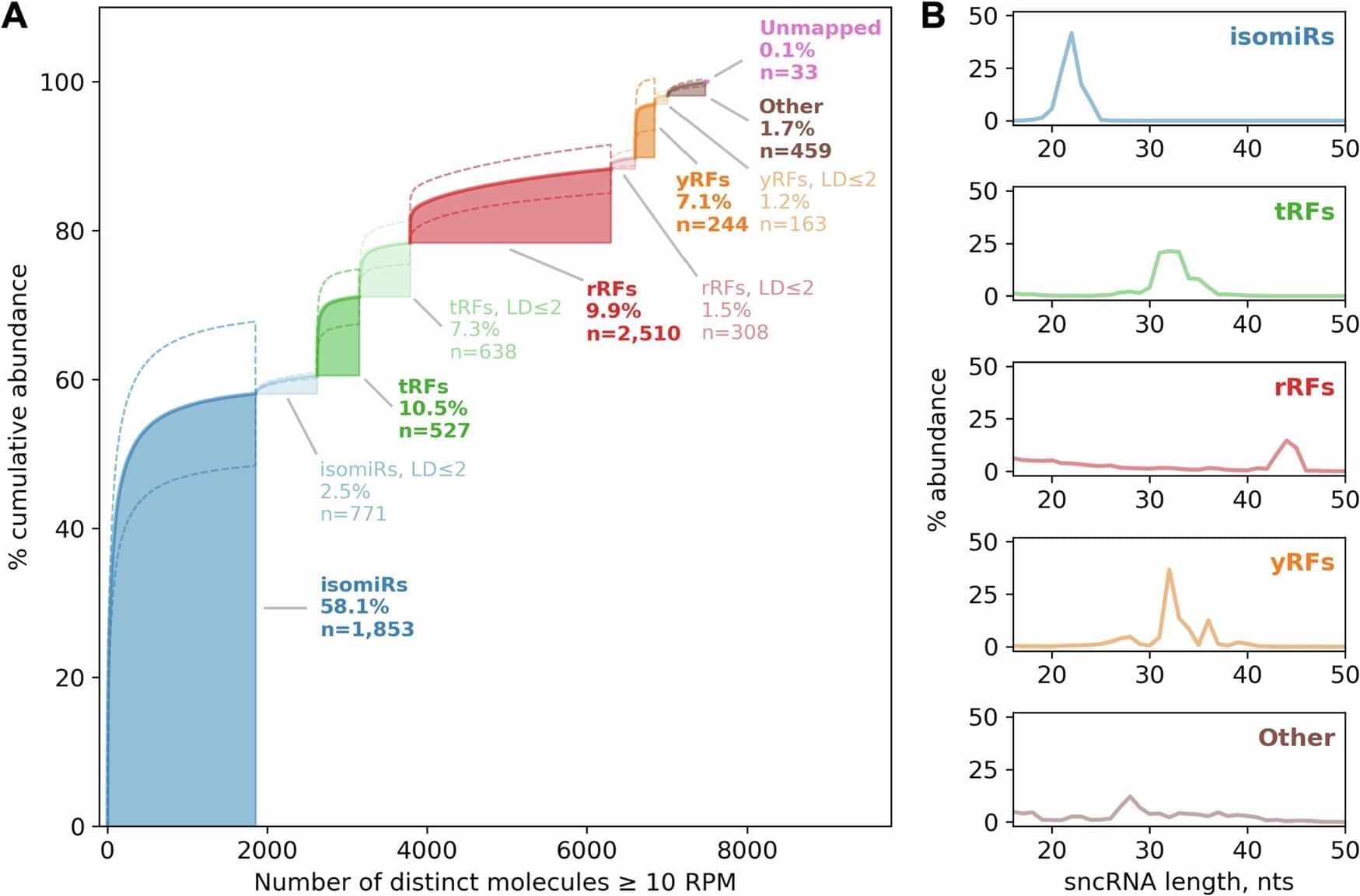
For decades, scientists studying brain disorders have focused almost exclusively on proteins and the genes encoding them. Now, research from Thomas Jefferson University’s Computational Medicine Center suggests that several classes of small regulatory molecules, fittingly known as small RNAs, may play a much larger role in schizophrenia and bipolar disorder, and in a healthy brain, than previously thought.
In a study recently published in Translational Psychiatry, a team led by Isidore Rigoutsos, Ph.D. took a comprehensive look at small RNAs in brain samples from people with schizophrenia, bipolar disorder and individuals without psychiatric illness. Their goal was to find out what kind of small RNAs are active in the brain, and whether their levels change in disease.
“Little attention had been paid to small RNAs in these disorders,” says Dr. Rigoutsos, “even though small RNAs help control numerous processes by modulating the abundance of genes.”

Researchers from Brown University and Mass General Brigham have developed an implantable brain-computer interface that allowed two people with paralysis — one with ALS and one with a spinal cord injury — to communicate through rapid, accurate typing. The system uses microelectrode sensors in the motor cortex, maps letters to attempted finger movements on a QWERTY keyboard, and decodes those neural signals into text.
In the study, one participant reached a top speed of 110 characters per minute (about 22 words per minute) with a 1.6% word error rate, and both participants were able to use the system from home after calibration with as few as 30 sentences. The results were published in Nature Neuroscience.
This is the kind of neurotechnology that starts to close the gap between thought and communication.
Implantable device research from the BrainGate clinical trial enables communication through rapid typing for a patient with ALS and a patient with a spinal cord injury.