A synaptic-resolution map of the neural circuits of a Drosophila larval brain reveals its connection types, neuron types, and circuit motifs.

I think an AI girlfriend or boyfriend would mess up your social skills and emotions/mental health. The article mentions other issues as well. It could turn some into “incels” meaning feeling resentful towards real women and possibly violent, not just domestic violence in my opinion but I’m not an expert but I’ve read about that a bit.
Chatbots such as Eva AI are getting better at mimicking human interaction but some fear they feed into unhealthy beliefs around gender-based control and violence.

This is true. I’ve been missing photography and books. Too much YouTube even can give you a bit of depression and anxiety. I’m a YouTube addict I’ll admit.
Reducing social media usage by as little as 15 minutes per day can increase health and well-being, claims a new study published in the Journal of Technology in Behavioral Science. The findings indicate that a 15-minute reduction in social media usage has positive consequences for one’s social life, vitality, and health.
Research has shown that excessive social media use can lead to a range of negative outcomes, including increased feelings of loneliness, anxiety, and depression. Social media use has also been linked to poor sleep quality, decreased physical activity, and decreased face-to-face social interaction. These negative outcomes are particularly concerning given that young adults are among the heaviest social media users, with many spending several hours daily on social media platforms.
Despite the growing concern about the negative effects of social media use, there is limited research on the effectiveness of interventions aimed at reducing social media use. Previous studies have suggested reducing social media use can improve mental health outcomes. However, small sample sizes and reliance on self-reported measures of social media use and mental health outcomes have limited these studies.
Autophagy biology has emerged as a ray of hope in addressing age-related diseases such as neurodegenerative disorders. Substantial effort in academia has been directed at advancing our understanding of the field and paving the way for ground-breaking therapies. But with genuine challenges in harnessing the power of autophagy and in developing effective therapies in this disease area, how close are we to really finding the first autophagy boosting drugs…?
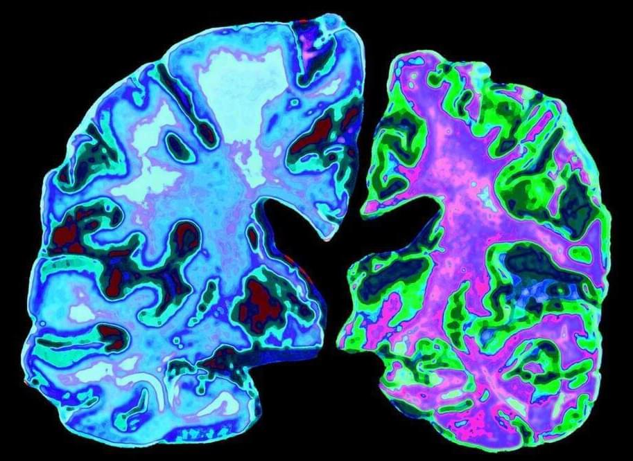
The devastating impact of neurodegenerative diseases such as Parkinson’s, Alzheimer’s and amyotrophic lateral sclerosis (ALS), the most common form of Motor Neurone Disease (MND), cannot be overstated. According to the WHO, neurological diseases affect over a billion people globally and are the leading cause of disability and the second leading cause of death worldwide [1, 2]. Incidence is increasing too, predominantly driven by population growth and aging. And, with no prospect of a cure, people who develop these conditions face a bleak future.
Justifiably, this disease area has been the subject of intensive research for many years and there have been some breakthroughs along the way, possibly offering hope for the development of new therapies. However, translating scientific breakthroughs into viable drugs for patients has been enormously challenging.
Neurologists at a memory clinic in China diagnosed a 19-year-old with what they believe to be Alzheimer’s disease, making him the youngest person to be diagnosed with the condition in the world.
The male teenager began experiencing memory decline around age 17, and the cognitive losses only worsened over the years.
Imaging of the patient’s brain showed shrinkage in the hippocampus, which is involved in memory, and his cerebrospinal fluid hinted at common markers of this most common form of dementia.
A protein fragment from the brain could be used to track progression of Alzheimer’s disease. The tell-tale fragment of tau protein was detectable in the cerebrospinal fluid (CSF) of patients with the disease, and researchers now hope to develop a blood test using it.
Alzheimer’s is a disease of errant protein aggregates that include amyloid plaques and then later tau tangles. ‘Amyloid plaques begin to form about 10 to 20 years before the first symptoms of Alzheimer’s,’ says Randall Bateman, a neurologist at Washington University School of Medicine who helped lead the study. ‘Whereas tau tangles begin once the symptoms begin.’
Studies in two patient groups, each with hundreds of people, revealed that levels of the fragment in the CSF were indicative of tau clumps in the brain seen on imaging and were linked to symptoms of cognitive decline.
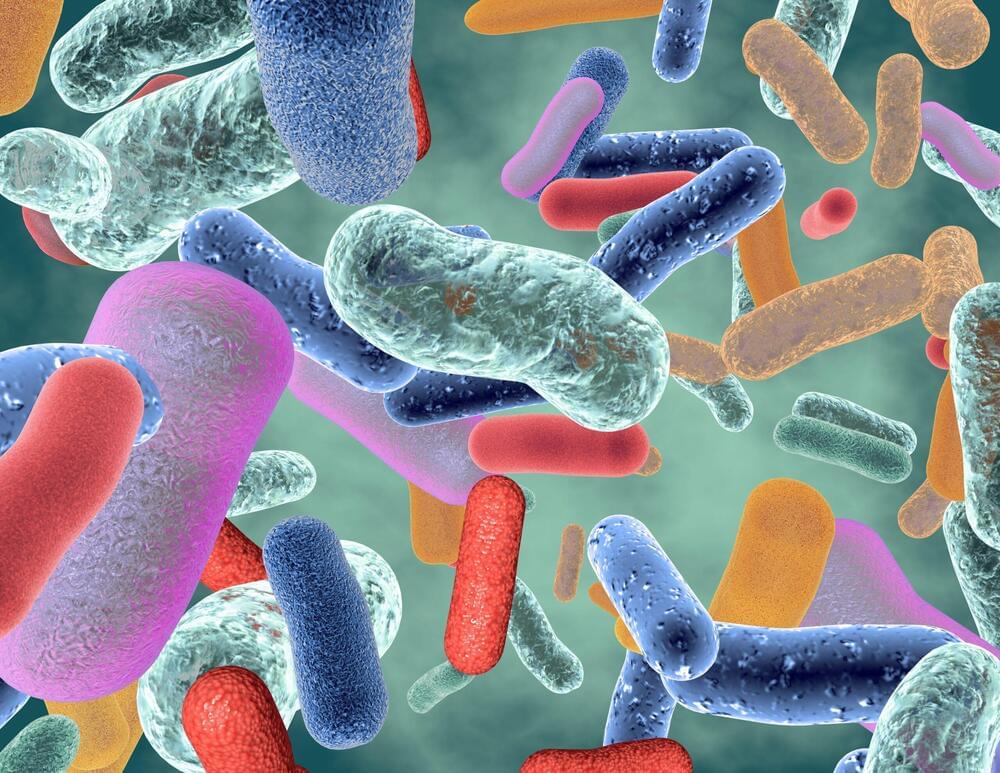
Our gut microorganisms, collectively known as the gut microbiota, affect far more than digestion. Bacteria in our intestinal tracts influence brain activity — and even the likelihood of developing mental disorders. Decades of research have shown that a bacterially imbalanced gut can disrupt many systems in the human body, contributing to obesity, malnutrition and even cancer. In a study published May 10 in Nature, Stanford Medicine researchers and collaborators used an ingestible device to capture the diversity of microorganisms, viruses, proteins and bile in the small intestine.
The proof-of-concept results provide early evidence that there are more comprehensive ways to measure microbiota in the digestive system than current sampling methods — which mostly focus on stool — and shed new light on how resident gut microbes might contribute to human physiology and disease.
“This paper demonstrates a big leap forward in microbial detection and captures the living gut microbiota in a nutshell,” said co-senior author KC Huang, PhD, a professor of bioengineering and of microbiology and immunology, co-senior author with David Relman, MD, a professor of medicine and of microbiology and immunology. “Samples from current tools don’t fully represent what’s going on inside of us. But it’s all we’ve had — until now.”
I gotta admit although effective and innovative, it’s also kinda creepy.
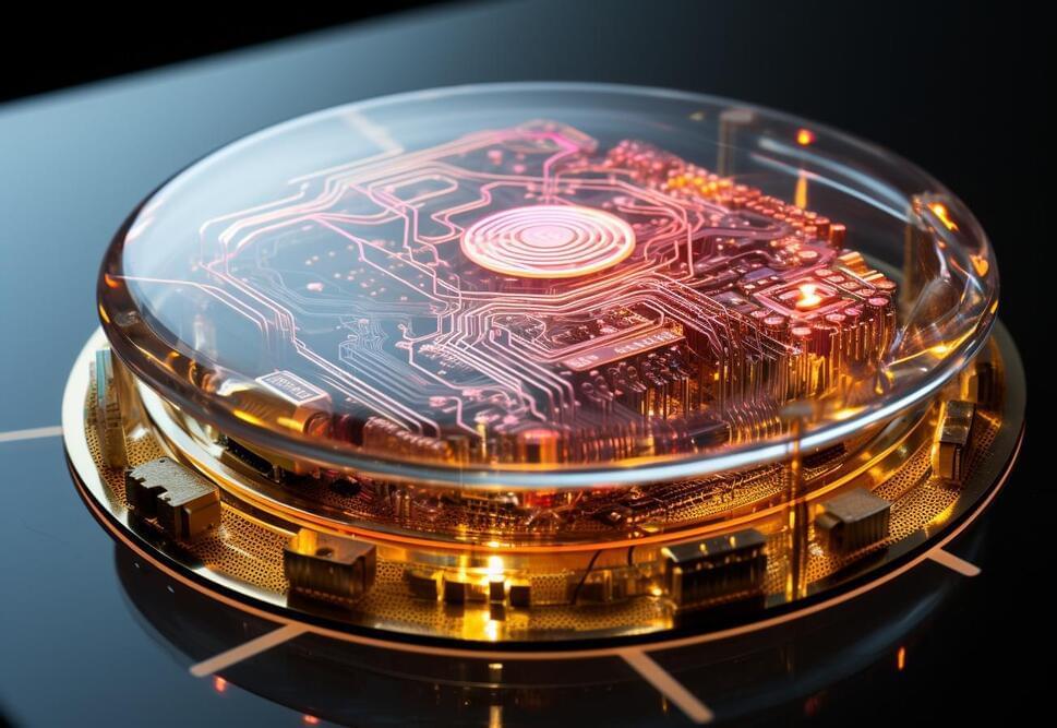
Last year, Monash University scientists created the “DishBrain” – a semi-biological computer chip with some 800,000 human and mouse brain cells lab-grown into its electrodes. Demonstrating something like sentience, it learned to play Pong within five minutes.
The micro-electrode array at the heart of the DishBrain was capable both of reading activity in the brain cells, and stimulating them with electrical signals, so the research team set up a version of Pong where the brain cells were fed a moving electrical stimulus to represent which side of the “screen” the ball was on, and how far away from the paddle it was. They allowed the brain cells to act on the paddle, moving it left and right.
Then they set up a very basic-reward system, using the fact that small clusters of brain cells tend to try to minimize unpredictability in their environment. So if the paddle hit the ball, the cells would receive a nice, predictable stimulus. But if it missed, the cells would get four seconds of totally unpredictable stimulation.