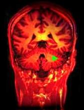
A recent study on monkeys found that stimulating a certain part of the forebrain wakes monkeys from anesthesia.

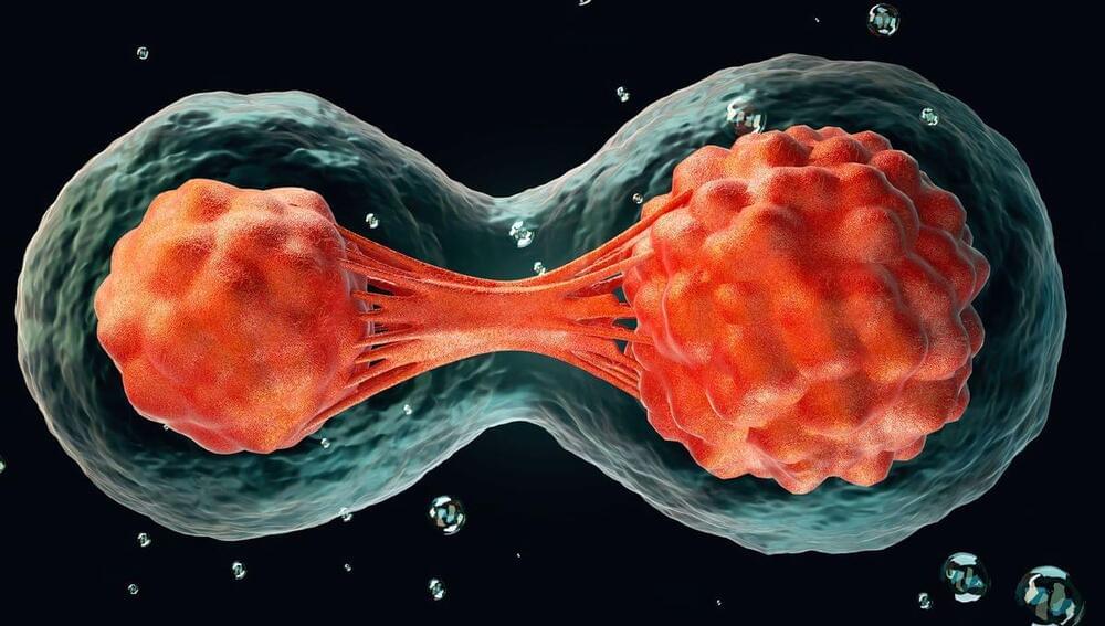
Summary: Treating a mouse model of multiple sclerosis (MS) with the pregnancy hormone estriol could reverse myelin breakdown in the brain’s cortex, a primary area affected in MS.
MS results in inflammation that damage the myelin coating around nerve fibers in the brain’s cortex, leading to disability worsening. Current MS treatments only target inflammation and can’t repair myelin damage.
However, the new study found that estriol not only prevented brain atrophy but also induced remyelination, suggesting it could repair MS-induced damage.
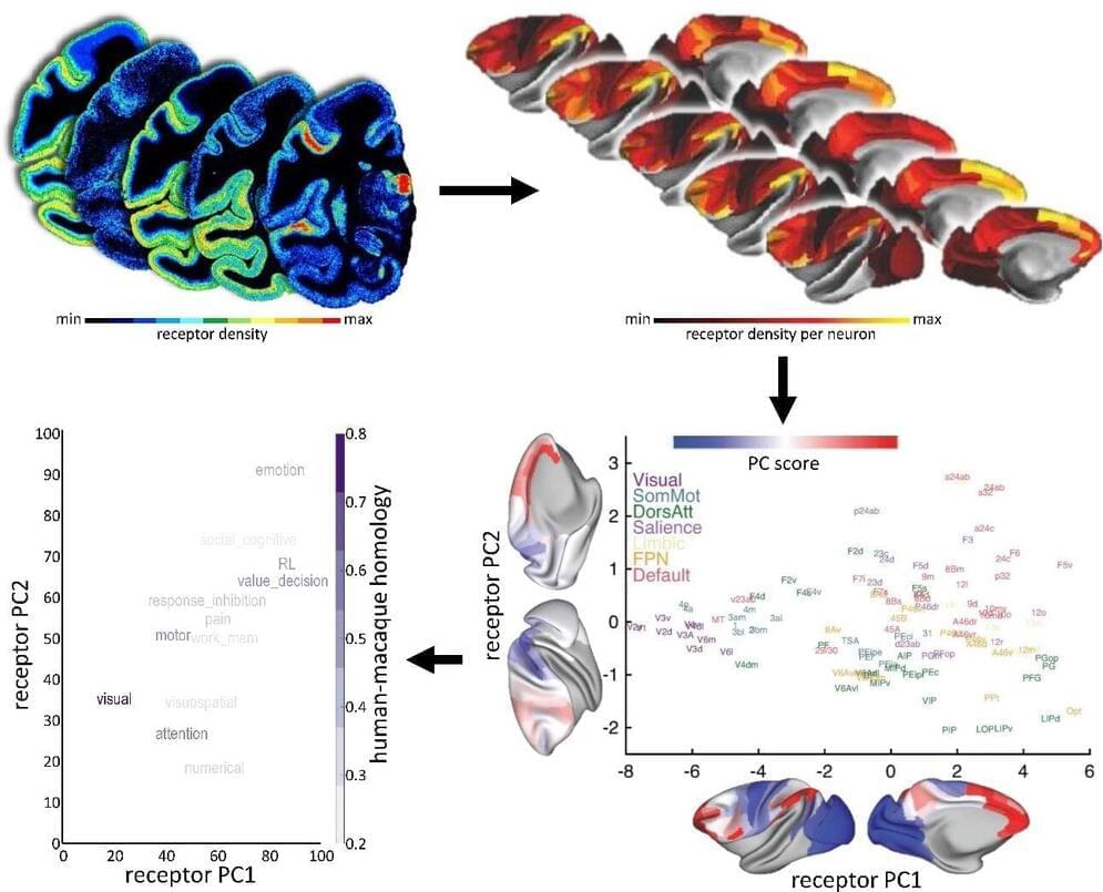
A key challenge in neuroscience is to understand how the brain can adapt to a changing world, even with a relatively static anatomy. The way the brain’s areas are structurally and functionally related to each other—its connectivity—is a key component. In order to explain its dynamics and functions, we also need to add another piece to the puzzle: receptors.
Now, a new mapping by Human Brain Project (HBP) researchers from the Forschungszentrum Jülich (Germany) and Heinrich-Heine-University Düsseldorf (Germany), in collaboration with scientists from the University of Bristol (UK), New York University (U.S.), Child Mind Institute (U.S.), and University of Paris Cité (France) had made advances on our understanding of the distribution of receptors across the brain.
The findings were published in Nature Neuroscience, and the data is now freely available to the neuroscientific community via the HBP’s EBRAINS infrastructure.

Nima Majlesi, director of Medical Toxicology at Staten Island University Hospital, also not part of the research, said the new study is “fascinating, and the more research that can be done on neurodegenerative diseases such as [Alzheimer’s disease], the more answers that can then be obtained for the betterment of everyone’s health.”
“There has never been any doubt that excessive alcohol use and recurrent intoxication [are] unhealthy in the medical community. There has occasionally been some doubt on whether a small amount of alcohol use daily can have health benefits. Even in patients not at risk for [Alzheimer’s disease], excessive alcohol use and recurrent intoxication [have] many detrimental effects on human health.” — Dr. Nima Majlesi
However, Dr. Majlesi cautioned that “in this study, they exposed mice to ethanol vapors, which is not the typical route for human consumption.”
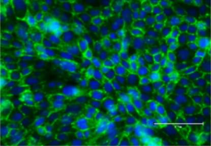
Scientists from the HIV Cure Center at the UNC School of Medicine, University of California San Diego, Emory University, and University of Pennsylvania have been searching for where exactly these latent cells are hiding in the body. New research published in the Journal of Clinical Investigations confirms that microglial cells – which are specialized immune cells with a decade-long lifespan in the brain – can serve as a stable viral reservoir for latent HIV.
Yuyang Tang, PhD, and Guochun Jiang, PhD, in the UNC School of Medicine extracted living brain tissue to conclude that specialized immune cells in the brain can harbor latent but replication-competent HIV.
As a part of its life cycle, the human immunodeficiency virus-1 (HIV) inserts a copy of its DNA into human immune cells. Some of these newly infected immune cells can then transition into a dormant, latent state for a long period of time, which is referred to as HIV latency.
Although current therapies, such current antiretroviral therapy (ART), can successfully block the virus from replicating further, it cannot eradicate latent HIV. If treatment is ever discontinued, the virus can rebound from latency and reignite the progression of HIV infection to AIDS.
Background
Many everyday tasks can fall under the mathematical class of “hard” problems. Typically, these problems belong to the complexity class of nondeterministic polynomial (NP) hard. These tasks require systematic approaches (algorithms) for optimal outcomes. In the case of significant complex problems (e.g., the number of ways to fix a product or the number of stops to be made on a delivery trip), more computations are required, which rapidly outgrows cognitive capacities.
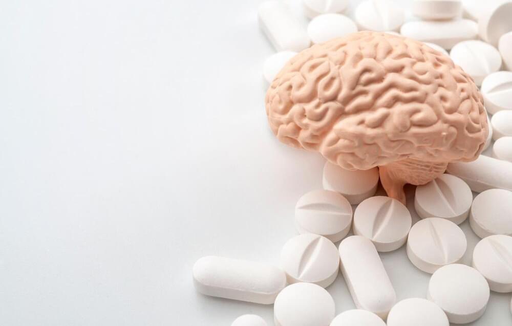
A recent Science Advances study investigated the effectiveness of three popular smart drugs, namely, modafinil (MOD), methylphenidate (MPH), and dextroamphetamine (DEX), against the difficulty of real-life daily tasks, i.e., the 0–1 knapsack optimization problem (“knapsack task”). A knapsack task is basically a combinatorial optimization task, the class of NP-time challenging problems.
Synthetic human embryos – derived from stem cells without the need for eggs or sperm – have been created for the first time, scientists say. The structures represent the very earliest stages of human development, which could allow for vital studies into disorders like recurrent miscarriage and genetic diseases. But questions have been posed about the legal and ethical implications, as the pace of scientific discovery outstrips the legislation.
The breakthrough was reported by the Guardian newspaper following an announcement by Professor Magdalena Żernicka-Goetz, a developmental biologist at the University of Cambridge and Caltech, at the 2023 annual meeting of the International Society for Stem Cell Research. The findings have not yet been published in a peer-reviewed paper.
It’s understood that the synthetic structures model the very beginnings of human development. They do not yet contain a brain or heart, for example, but comprise the cells that would be needed to form a placenta, yolk sac, and embryo. Żernicka-Goetz told the conference that the structures have been grown to just beyond the equivalent of 14 days of natural gestation for a human embryo in the womb. It’s not clear whether it would be possible to allow them to mature any further.
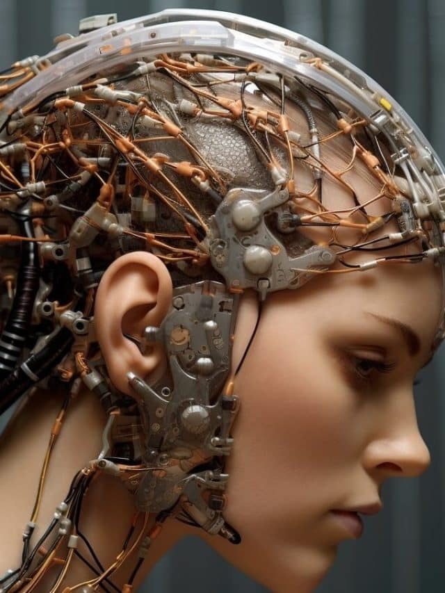

The brain is regarded as one of the most complex known structures in the universe. It has billions of neurons, trillions of connections, and multiple levels ranging from cellular to molecular and synaptic. But the biggest challenge is that the brain is difficult to access.
“The brain is encased in a thick bone,” said Kinney, “and if you try to access, poke, or prod it, it will get really upset and hemorrhage, and delicate neurons will die.”
Nevertheless, Kinney said progress is being made on various fronts, particularly in the field of recording brain activity, which is good news for those trying to build brain-like computers.
A new study published in Psychological Science investigated the relationship between loneliness, brain activity, and social interactions. The results suggest that individuals who experience loneliness may process social information differently from those who do not, contributing to feelings of isolation and disconnection.
The study highlights the importance of social connection for psychological well-being. It emphasizes the need for further research in this area to develop effective interventions to help individuals experiencing loneliness improve their social connections and overall quality of life.
Humans are social creatures, and social connection is essential for physical and mental health. Social isolation and loneliness have been linked to various adverse outcomes, including depression, anxiety, cardiovascular disease, and even mortality.