Toggle light / dark theme

Scientists become research subjects in after-hours brain-scanning project

A quest to analyze the unique features of individual human brains evolved into the so-called Midnight Scan Club, a group of scientists who had big ideas but almost no funding and little time to research the trillions of neural connections that activate the body’s most powerful organ.

The research group started in 2013 by two neuroscientists at Washington University School of Medicine in St. Louis who aimed to collect a massive amount of data on individual brains. The study’s subjects were the scientists themselves and eight others, all junior faculty or graduate students.

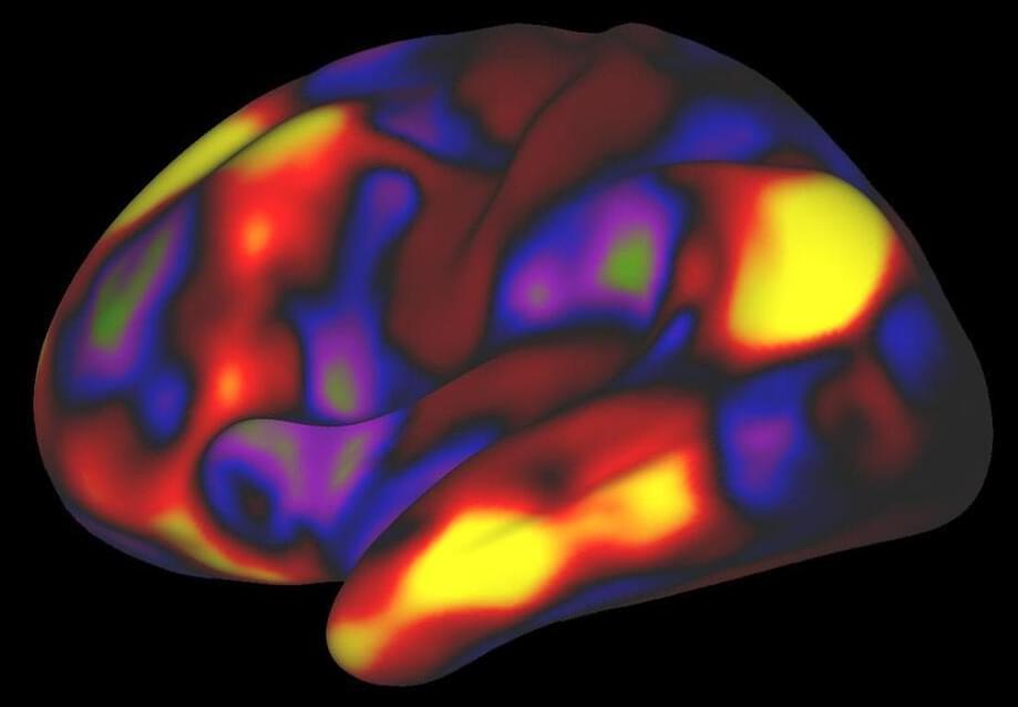
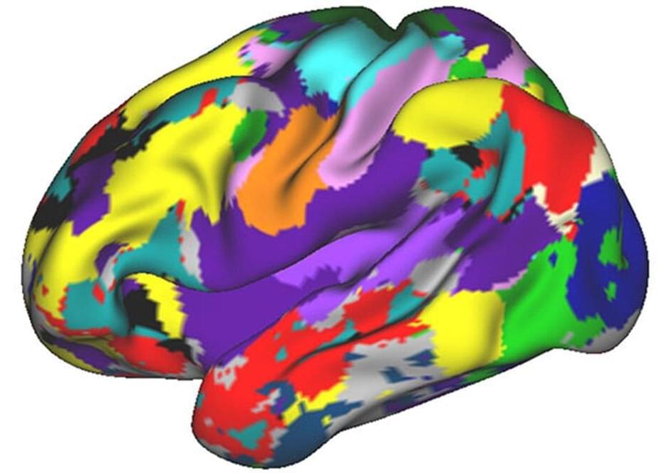
Most efforts to analyze connections involve scanning many brains and averaging the data across groups of people. For this study, the researchers used brain-imaging techniques to evaluate brain networks that control speech and motor function, among other activities. The researchers examined individuals while resting and performing cognitive tasks such as reading.

Researchers discover new information about the effects of sleep on the human brain

The Neuro-Network.

𝐑𝐞𝐬𝐞𝐚𝐫𝐜𝐡𝐞𝐫𝐬 𝐝𝐢𝐬𝐜𝐨𝐯𝐞𝐫 𝐧𝐞𝐰 𝐢𝐧𝐟𝐨𝐫𝐦𝐚𝐭𝐢𝐨𝐧 𝐚𝐛𝐨𝐮𝐭 𝐭𝐡𝐞 𝐞𝐟𝐟𝐞𝐜𝐭𝐬 𝐨𝐟 𝐬𝐥𝐞𝐞𝐩 𝐨𝐧 𝐭𝐡𝐞 𝐡𝐮𝐦𝐚𝐧 𝐛𝐫𝐚𝐢𝐧

𝙐𝙣𝙞𝙫𝙚𝙧𝙨𝙞𝙩𝙮 𝙤𝙛 𝙊𝙪𝙡𝙪 𝙁𝙪𝙣𝙘𝙩𝙞𝙤𝙣𝙖𝙡 𝙉𝙚𝙪𝙧𝙤𝙞𝙢𝙖𝙜𝙞𝙣𝙜 𝙧𝙚𝙨𝙚𝙖𝙧𝙘𝙝 𝙜𝙧𝙤𝙪𝙥 𝙝𝙖𝙨 𝙛𝙤𝙧 𝙩𝙝𝙚 𝙛𝙞𝙧𝙨𝙩 𝙩… See more.


The University of Oulu Functional Neuroimaging research group has for the first time succeeded in describing how the various types of pulsations in the human brain change when a person sleeps.

Risks of mental health outcomes in people with covid-19: cohort study

Objective To estimate the risks of incident mental health disorders in survivors of the acute phase of covid-19.

Design Cohort study.

Setting US department of veterans affairs.

Participants Cohort comprising 153 848 people who survived the first 30 days of SARS-CoV-2 infection, and two control groups: a contemporary group (n=5 637 840) with no evidence of SARS-CoV-2, and a historical control group (n=5 859 251) that predated the covid-19 pandemic.

Depression and Alzheimer’s Disease Share Common Genetic Roots

Epidemiological data have long linked depression with Alzheimer’s disease (AD), a neurodegenerative disease characterized by progressive dementia that affects nearly 6 million Americans. Now, a new study identifies common genetic factors in both depression and AD. Importantly, the researchers found that depression played a causal role in AD development, and those with worse depression experienced a faster decline in memory. The study appears in Biological Psychiatry, published by Elsevier.

Co-senior author Aliza Wingo, MD, of Emory University School of Medicine, Atlanta, USA, said of the work, “It raises the possibility that there are genes that contribute to both illnesses. While the shared genetic basis is small, the findings suggest a potential causal role of depression on dementia.”

The authors performed a genome-wide association study (GWAS), a technique that scans the entire genome for areas of commonality associated with particular conditions. The GWAS identified 28 brain proteins and 75 transcripts – the messages that encode proteins – that were associated with depression. Among those, 46 transcripts and 7 proteins were also associated with symptoms of AD. The data suggest a shared genetic basis for the two diseases, which may drive the increased risk for AD associated with depression.

Study shows that individual neurons could learn

Humans have been trying to understand how the brain works and how it acquires information for centuries. While neuroscientists now have a pretty good understanding of how different parts of the brain work and what their function is, many questions remain unanswered; thus, a unified neuroscience theory is still lacking.

In recent years, computer scientists have been trying to create computational tools that artificially recreate the functions and processes of the human . New theories clarifying how the brain makes predictions could help to significantly enhance these tools so that they replicate neural functions in increasingly realistic ways.

Researchers at the Canadian Centre for Behavioural Neuroscience in Lethbridge, Canada have recently carried out a study investigating how individual learn and make predictions about the future. Their findings, published in Nature Machine Intelligence, suggest that the ability of single neurons to predict their future activity could offer a new learning mechanism.

Touch Sensitive Brain Cells Controlled by Micromagnets

Summary: A newly developed technique allows researchers to remotely active neurons with the aid of microscopic magnetic particles.

Source: UCL

Scientists at UCL have developed a new technique that uses microscopic magnetic particles to remotely activate brain cells; researchers say the discovery in rats could potentially lead to the development of a new class of non-invasive therapies for neurological disorders.

New project creates digital clones of human brains to help treat neurological disorders

More recently, digital twins have been the focus of a European Union-funded project that seeks to clone a patient’s entire brain. Dubbed Neurotwin, the research project aims to create virtual models that can be used to predict the effects of stimulation for the treatment of neurological disorders—including epilepsy and Alzheimer’s disease. When it comes to epilepsy, non-invasive stimulations (where electrical currents are painlessly delivered to the brain) have proven effective in tackling seizures. Given how drugs don’t help a third of epilepsy patients, the technology is coveted yet needs refinement. This is where virtual clones come in.

“The digital avatar is essentially a mathematical model running on a computer,” Giulio Ruffini, coordinator of the Neurotwin project, told WIRED. Including a network of embedded “neural mass models,” the technology hopes to create a map of the neural connections in the brain—a concept termed as the ‘connectome’. “In the case of epilepsy, some areas of the connectome could become overexcited,” the outlet mentioned. “In the case of, say, stroke, the connectome might be altered.” Once the digital clone has been created by the team, with about half an hour-worth of magnetic resonance imaging (MRI) data and ten minutes of electroencephalography (EEG) readings to capture electrical activities and realistically simulate the brain’s main tissues (including the scalp, skull, cerebrospinal fluid, and grey and white matter), it can then be used to optimise stimulation of the real patient’s brain.

According to Ruffini, this is possible “because we can run endless simulations on the computer until we find what we need. It is, in this sense, like a weather forecasting computational model.”

Better understanding communication between neurons in the brain

In the field of optogenetics, scientists investigate the activity of neurons in the brain using light. A team led by Prof. Dr. Ilka Diester and Dr. David Eriksson from the Optophysiology Laboratory at the University of Freiburg has developed a new method to simultaneously conduct laminar recordings, multifiber stimulations, 3D optogenetic stimulation, connectivity inference, and behavioral quantification on brains. Their results are presented in Nature Communications. “Our work paves the way for large-scale photo-recording and controlled interrogation of fast neural communication in any combination of brain areas,” Diester explains. “This can help us unravel the rapid and multilayered dialogs between neurons that maintain brain function.”

The research group, in collaboration with Dr. Patrick Ruther of the Department of Microsystems Engineering (IMTEK) at the University of Freiburg, is developing a new method for the controlled interrogation and recording of neuronal activity in the . To do this, the team is taking advantage of thin, cell-sized optical fibers for minimally invasive optogenetic implantation. “We combine side-emitting fibers with silicon probes to achieve high-quality recordings and ultrafast, multichannel optogenetic control.”

They call the system Fused Fiber Light Emission and eXtracellular Recording, or FFLEXR. In addition to optical fibers that can be attached to any silicon probe, the uses linear depth-resolved , a lightweight fiber matrix connector, a flexible multifiber ribbon cable, an optical commutator for efficient multichannel stimulation, a general-purpose patch cable, and an algorithm to manage the photovoltaic response.

/* */