In this episode, I interview Dr. Robert Sapolsky, Ph.D., Professor of Biology, Neurology & Neurosurgery at Stanford University. We discuss stress, what defines short-term versus long-term stress, and how stress can be beneficial or detrimental, depending on the context. We also discuss stress mitigation and how our sense of control over stress mitigation techniques, including exercise, determine health outcomes. Dr. Sapolsky explains some of the key effects of the hormone testosterone — how it can amplify pre-existing tendencies for aggression or sexual behavior, but that it does not produce those behaviors per se. He also explains how testosterone impacts our social hierarchies, sense of confidence, and willingness to embrace challenges of different kinds. He also explains how our behaviors and perceptions shape testosterone levels. And we discuss estrogen and the powerful role it plays in brain development, health and longevity. Finally, we discuss free will, what it means to have free will, and if we have any free will, including how knowledge alone might allow us to make better decisions for ourselves and society.
Davis (2014) called for “extreme caution” in the use of non-invasive brain stimulation (NIBS) to treat neurological disorders in children, due to gaps in scientific knowledge. We are sympathetic to his position. However, we must also address the ethical implications of applying this technology to minors. Compensatory trade-offs associated with NIBS present a challenge to its use in children, insofar as these trade-offs have the effect of limiting the child’s future options. The distinction between treatment and enhancement has some normative force here. As the intervention moves away from being a treatment toward being an enhancement—and thus toward a more uncertain weighing of the benefits, risks, and costs—considerations of the child’s best interests (as judged by the parents) diminish, and the need to protect the child’s (future) autonomy looms larger. NIBS for enhancement involving trade-offs should therefore be delayed, if possible, until the child reaches a state of maturity and can make an informed, personal decision. NIBS for treatment, by contrast, is permissible insofar as it can be shown to be at least as safe and effective as currently approved treatments, which are (themselves) justified on a best interests standard.
Exercise can have surprisingly transformative impacts on the brain, according neuroscientist Wendy Suzuki. It has the power not only to boost mood and focus due to the increase in neurotransmitters like dopamine, serotonin, and noradrenaline, but also contributes to long-term brain health. Exercise stimulates the growth of new brain cells, particularly in the hippocampus, improving long-term memory and increasing its volume. Suzuki notes that you don’t have to become a marathon runner to obtain these benefits — even just 10 minutes of walking per day can have noticeable benefits. It just takes a bit of willpower and experimentation.
0:00 My exercise epiphany. 1:35 What is “runner’s high”? 2:40 The hippocampus & prefrontal cortex. 3:32 Neuroplasticity: It’s never too late to move your body.
How can consciousness be addressed scientifically? The Tucson conference, founded in 1994 and celebrating its 20th anniversary in 2014, exemplifies the quest. What are the range of theories? Where do participants position themselves? Meet the founders, early visionaries, new scientists and thinkers. Progress is being made, but what does this really mean?
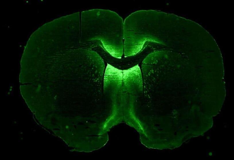
Fragile X syndrome is a genetic disorder caused by a mutation in a gene that lies at the tip of the X chromosome. It is linked to autism spectrum disorders.
People with fragile X experience a range of symptoms that include cognitive impairment, developmental and speech delays and hyperactivity. They may also have some physical features such as large ears and foreheads, flabby muscles and poor coordination.
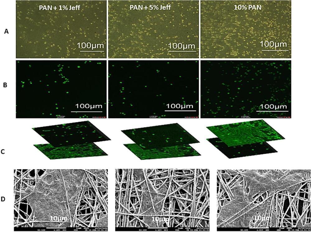
Scientists have found a way to use nanotechnology to create a 3D “scaffold” to grow cells from the retina—paving the way for potential new ways of treating a common cause of blindness.
Researchers, led by Professor Barbara Pierscionek from Anglia Ruskin University (ARU), have been working on a way to successfully grow retinal pigment epithelial (RPE) cells that stay healthy and viable for up to 150 days. RPE cells sit just outside the neural part of the retina, and when damaged, can cause vision to deteriorate. Their work is published in Materials & Design.
It is the first time this technology, called “electrospinning,” has been used to create a scaffold on which the RPE cells could grow, and could revolutionize treatment for one of age-related macular degeneration, one of the world’s most common vision complaints.
For many people, depression turns out to be one of the most disabling illnesses that we have in society. Despite the treatments that we have available, many people are not responding that well. It’s a disorder that can be very disabling in society. It’s also a disorder that has medical consequences. By understand the neurobiology of depression we hope to be able more to find the right treatment for the patient suffering from this disease. The current standard of care for the treatment of depression is based on what we call the monoamine deficiency hypothesis. Essentially, presuming that one of three neurotransmitters in the brain is deficient or underactive. But the reality is, there are more than 100 neurotransmitters in the brain. And billions of connections between neurons. So we know that that’s a limited hypothesis. Neurotransmitters can be thought of as the chemical messengers within the brain, it’s what helps one cell in the brain communicate with another, to pass that message along from one brain region to another. For decades, we thought that the primary pathology, the primary cause of depression was some abnormality in these neurotransmitters, specifically serotonin or norepinephrine. However, norepinephrine and serotonin did not seem to be able to account for this cause, or to cause the symptoms of depression in people who had major depression. Instead, the chemical messengers between the nerve cells in the higher centers of the brain, which include glutamate and GABA, were possibilities as alternative causes for the symptoms of depression. When you’re exposed to severe and chronic stress like people experience when they have depression, you lose some of the connections between the nerve cells. The communication in these circuits becomes inefficient and noisy, we think that the loss of these synaptic connections contributes to the biology of depression. There are clear differences between a healthy brain and a depressed brain. And the exciting thing is, when you treat that depression effectively, the brain goes back to looking like a healthy brain, both at the cellular level and at a global scale. It’s critical to understand the neurobiology of depression and how the brain plays a role in that for two main reasons. One, it helps us understand how the disease develops and progresses, and we can start to target treatments based on that. We are in a new era of psychiatry. This is a paradigm shift, away from a model of monoaminergic deficiency to a fuller understanding of the brain as a complex neurochemical organ. All of the research is driven by the imperative to alleviate human suffering. Depression is one of the most substantial contributors to human suffering. The opportunity to make even a tiny dent in that is an incredible opportunity.
Can the fountain of youth come in the form a pill?
Imagine this: a cocktail of specialized chemicals that rejuvenates your whole body, from your eyes and brain to your kidneys and muscles—bringing you back to a more youthful version of yourself.