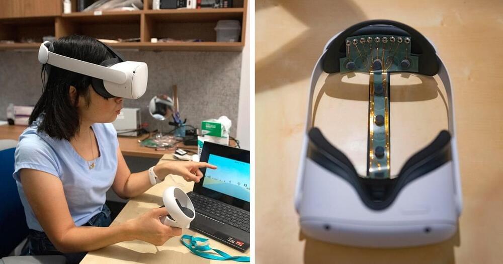
Toggle light / dark theme

Monogamous prairie voles form lifelong pair bonds, but the neuronal dynamics that underlie bond formation and maintenance in this species remain largely unknown. We performed imaging of populations of neurons while voles interacted with their pair-bonded partner or a novel vole before and after bond formation. We identified neurons that were active during partner approach and found that this subset of cells was distinct from those that were active during novel approach. Furthermore, the number of partner approach cells increased following bond formation, reflecting the emergence of bonding behaviors and correlating with bond strength. This discovery sheds light on how pair bonds may be encoded within the brain and what changes as bonds mature.

Pair-bond formation depends vitally on neuromodulatory signaling within the nucleus accumbens, but the neuronal dynamics underlying this behavior remain unclear. Using 1-photon in vivo Ca2+ imaging in monogamous prairie voles, we found that pair bonding does not elicit differences in overall nucleus accumbens Ca2+ activity. Instead, we identified distinct ensembles of neurons in this region that are recruited during approach to either a partner or a novel vole. The partner-approach neuronal ensemble increased in size following bond formation, and differences in the size of approach ensembles for partner and novel voles predict bond strength. In contrast, neurons comprising departure ensembles do not change over time and are not correlated with bond strength, indicating that ensemble plasticity is specific to partner approach.

During sleep and awake rest, the neocortex generates large-scale slow-wave (SW) activity. Here, we report that the claustrum coordinates neocortical SW generation. We established a transgenic mouse line that enabled the genetic interrogation of a subpopulation of claustral glutamatergic neurons. These neurons received inputs from and sent outputs to widespread neocortical areas. The claustral neuronal firings mostly correlated with cortical SW activity. In vitro optogenetic stimulation of the claustrum induced excitatory postsynaptic responses in most neocortical neurons, but elicited action potentials primarily in inhibitory interneurons. In vivo optogenetic stimulation induced a synchronized down-state featuring prolonged silencing of neural activity in all layers of many cortical areas, followed by a down-to-up state transition. In contrast, genetic ablation of claustral neurons attenuated SW activity in the frontal cortex. These results demonstrate a crucial role of claustral neurons in synchronizing inhibitory interneurons across wide cortical areas for the spatiotemporal coordination of SW activity.

Scientists have discovered a new treatment to dramatically reduce swelling after brain and spinal cord injuries, offering hope to 75 million victims worldwide each year.

The breakthrough in treating such injuries—referred to as central nervous system (CNS) edema—is thought to be hugely significant because current options are limited to putting patients in an induced coma or performing risky surgery.

Brain and affect all age groups. Older people are more at risk of sustaining them from strokes or falls, while for younger age groups, major causes include road traffic accidents and injuries from sports such as rugby, US-style football and other contact games.

For Army scientists, the goal of neuroscience research is pursuing the inner workings of the human brain to advance scientific understanding and improve Soldier performance.

Researchers recently applied new techniques to modify brain activity. Not only are these techniques used to characterize and study complex networks such as in telecommunications or social networks—they describe how different nodes, or elements of the network: brain regions in neuroscience, or individuals in social networks, interact with each other.

The U.S. Army Combat Capabilities Development Command’s Army Research Laboratory, in collaboration with academic partners, collaborated on a neurostimulation study, where they safely and non-invasively modified and then characterized the dynamics of the brain’s response to this modification. This research provides some of the foundational knowledge for future technologies that may one day expedite cognitive processes. The journal Network Neuroscience published the recent discoveries.

According to Li and Spitzer, running on a treadmill, or performing another sustained aerobic exercise—like dancing or kickboxing—on a regular basis might actually enhance motor skill-based learning.

When comparing the brains of mice that exercised versus those who did not, Li and Spitzer found that specific neurons switched their chemical signals (neurotransmitters), after exercising, which led to improved learning for motor skill-specific acquisition.

While physical exercise is proven to promote motor skill learning in normal individuals as well as those with neurological disorders, the mechanism of action is unclear. The study found that that one just week of voluntary wheel running enhances the acquisition of motor skills in normal adult mice. Voluntary being the keyword here.

Researchers at the RIKEN Center for Biosystems Dynamics Research (BDR) in Japan have identified changes in the aging brain related to blood circulation. Published in the scientific journal Brain, the study found that natural age-related enlargement of the ventricles—a condition called ventriculomegaly—was associated with a lag in blood drainage from a specific deep region of the brain. The lag can be detected easily with MRI, making it a potential biomarker for predicting ventriculomegaly and the aging brain, which can then be treated quickly.

Ventriculomegaly is an abnormal condition in which fluid accumulates in the ventricles of the without properly draining, making them enlarged. Although ventricular enlargement within normal range is not itself considered a disease, when left unchecked it can lead to ventriculomegaly and dementia resulting from normal pressure hydrocephalus. In their study, the team found that ventriculomegaly was associated with changes in circulation of the brain. “We found an age-related perfusion timing shift in the brain’s venous systems whose lifespan profile was very similar to, but slightly preceded that of ventricular enlargement,” explains first author Toshihiko Aso.

After blood circulates through the brain providing necessary oxygen, the deoxygenated blood must return to the heart though our veins. This happens through two pathways, one draining blood from regions close to the surface of the brain, and the other from areas deep in the brain. By using MRI to measure changes in , the team at BDR recently found that as we age, the time it takes for blood to drain through these two pathways becomes out of sync. The result is a time lag between the deep drainage and the surface pathway, which increases with age.