This is the third part in a series on deep brain stimulation for depression. Read from the beginning.
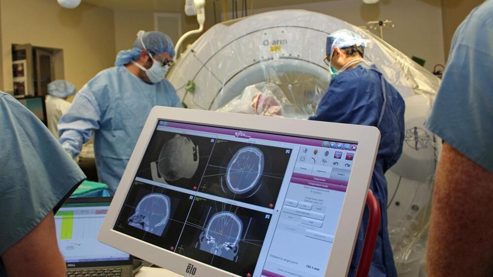
Early in the morning on August 22, 2022, Jon’s medical team told him that neurologist Helen Mayberg would check in with him just before his deep brain stimulation surgery.
This isn’t good. I feel for anyone in the human trials.
Documents viewed as part of a new investigation by Wired, however, as well as testimony from a former employee, contradict Musk’s claims entirely — and the details are as upsetting as they are damning, adding to a mounting case against the safety of Neuralink’s devices.
Here’s the harrowing casualty report, per veterinary records obtained by Wired from the California National Primate Research Center (CNPRC) at UC Davis, the site of the Neuralink primate research. Up to a dozen monkeys suffered grisly fates after receiving a Neuralink implant, including brain swelling and partial paralysis.
The types of cancer that occur in children often are different from those in adults. Childhood cancers usually are not linked to lifestyle or environmental risk factors, as is often the case in adults. Nonetheless, cancer is the second-leading cause of death in children 1 to 14 years old, according to the American Cancer Society. Nearly 10,000 children in the U.S. under the age of 15 will be diagnosed with cancer in 2023, and about 1,000 children are expected to die of the disease.
September is Childhood Cancer Awareness Month, which makes this a good time to learn about three of the most common types of cancer in children: acute lymphocytic leukemia, neuroblastoma and pediatric brain tumors.
Acute lymphocytic leukemia is a cancer of the blood and bone marrow. It’s the most common type of cancer in children, and treatments result in a good chance for a cure. Acute lymphocytic leukemia also can occur in adults, though the chance of a cure is greatly reduced.
Jellyfish are more advanced than once thought. A new study from the University of Copenhagen has demonstrated that Caribbean box jellyfish can learn at a much more complex level than ever imagined—despite only having one thousand nerve cells and no centralized brain. The finding changes our fundamental understanding of the brain and could enlighten us about our own mysterious brains.
After more than 500 million years on Earth, the immense evolutionary success of jellyfish is undeniable. Still, we’ve always thought of them as simple creatures with very limited learning abilities.
The prevailing opinion is that more advanced nervous systems equate with more advanced learning potential in animals. Jellyfish and their relatives, collectively known as cnidarians, are considered to be the earliest living animals to develop nervous systems and to have fairly simple nervous systems and no centralized brain.
Discovering And Developing Medicines To Keep You Biologically Young — Dr. Marco Quarta, Ph.D. — Co-Founder and CEO, Rubedo Life Sciences; CEO, Phaedon Institute.
Dr. Marco Quarta, Ph.D. is Co-Founder and CEO of Rubedo Life Sciences (https://www.rubedolife.com/), a biopharmaceutical company developing a broad portfolio of innovative therapies engineered to target cells which drive chronic age-related diseases. The company’s proprietary ALEMBIC™ drug discovery platform has engineered novel first-in-class small molecules designed to selectively target senescent cells, which play a key role in the progression of pulmonary, dermatological, oncological, neurodegenerative, fibrotic and other chronic disorders.
Dr. Quarta received his doctorate degree in Biotechnology from the University of Bologna and a Ph.D. in Neuroscience from the University of Padua. He completed a post-doc in Aging and Stem cell Biology in the lab of Prof. Thomas Rando at Stanford University and continued his work at Stanford directing a research team at the Center for Tissue Regeneration, Repair, and Restoration at the VA Hospital in Palo Alto, CA. While there, he established a translational program in regenerative medicine. He has over 35 publications and patents in the field of aging, stem cells, regenerative medicine, and rejuvenation.
Dr. Quarta also co-founded Wetware Concepts, Young European Biotech Network (YEBN), and Turn Biotechnology, and served as an executive board member of the European Federation of Biotechnologies. He currently sits on the advisory board of the California Institute for Regenerative Medicine (CIRM) Calpoly Bridge program, and the advisory board at the Center for Healthcare Innovation. He is a member of the Paul F Glenn Center for the Biology of Aging Studies at Stanford University, one of the most prestigious institutions supporting the science of aging.
Dr. Quarta also serves as CEO and President for the Board of Directors of The Phaedon Institute (https://www.phaedon.institute/), a think-tank organization that operates with the mission of supporting and enabling effective and sustainable growth in the field of aging and longevity sciences.
This November, researchers, clinicians, and investors will descend on Miami, Florida for the annual Wonderland conference. This year, the world’s leading psychedelics conference is expanding its focus to include longevity for the first time, welcoming top speakers from across the field, from Bryan Johnson to Aubrey de Grey.
Through a series of keynotes, round table and panel discussions, and town hall open mic sessions, the event aims to explore the increasingly linked topics of psychedelic medicine, mental health, and longevity medicine.
Longevity. Technology: Every month it seems, more and more research is highlighting the connection between mental health and longevity – from accelerated biological aging to reduced life expectancy. With psychedelics simultaneously demonstrating compelling results in the treatment of mental conditions, from depression to PTSD, the synergies between longevity and psychedelic medicine are clear. We caught up with leading longevity physician Dr Halland Chen to tap into his views on recent developments in longevity medicine and its links with the psychedelic world.
Stanislaw Ulam Memorial Lecture Series Ricard Solé Universitat Pompeu Fabra, SFI Lecture 2: Synthetic Brains and Minds: What is Possible? In our search fo…
As befits the child of a scientist, Martin Picard’s young son, 3, is already learning about biology with an age-appropriate textbook, “Cell Biology for Babies.” Picard winces a little whenever the book calls mitochondria the “powerhouses of the cell” but figures he has plenty of time as his son grows older to explain why the tiny organelles are much more than simple energy sources.
Picard is a leading proponent of mitochondrial psychobiology (a phrase he coined), an emerging field that examines how psychological states like stress influence mitochondrial functions, which in turn influence mental and physical health.
“The powerhouse analogy is outdated and one-dimensional and can impede science by limiting researchers’ perceptions of what mitochondria can do,” says Picard, associate professor of behavioral medicine in psychiatry and neurology.