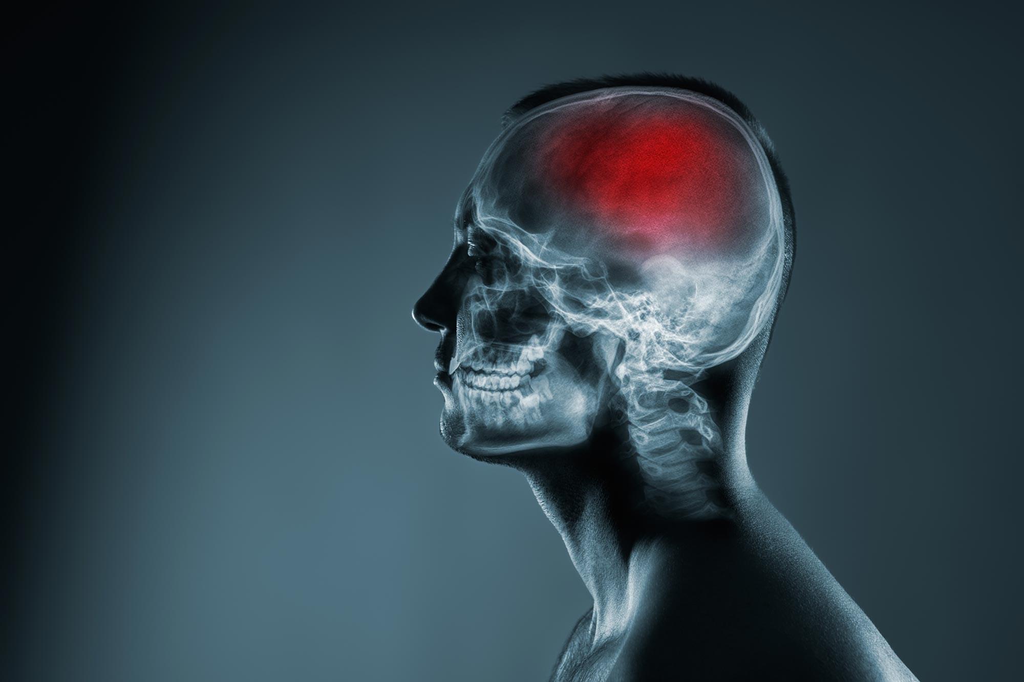
Lacunar stroke may arise from damage in the brain’s small blood vessels rather than fatty plaque in larger arteries, pointing researchers toward new treatment strategies.
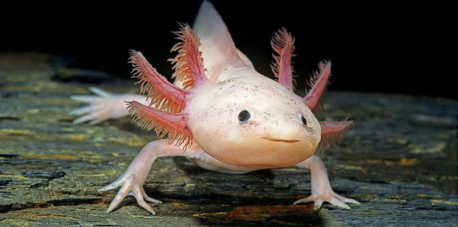
But over evolutionary time, mammals have obviously lost the vast majority of this regenerative capacity. Instead, evolution opted for faster wound sealing, stronger immune responses and more stable neural systems in mammals. This is likely because surviving injury would have mattered more than perfectly reconstructing tissue months later.
Salamanders, on the other hand, have retained far more of this ancestral regenerative toolkit. Their ecology may have reinforced this retention, since small amphibians are especially vulnerable to predation and environmental injury. Limbs, tails and nervous tissue can be damaged surprisingly easily in aquatic habitats filled with predators, debris, and competition. For an animal living close to the edge of survival, the ability to recover from catastrophic injury could dramatically improve reproductive success.
The axolotl’s strange life history has most probably also enabled this unique ability. Unlike many amphibians, axolotls remain in a juvenile-like aquatic state throughout adulthood, a phenomenon known as “neoteny.” Intriguingly, juvenile tissues in many vertebrates tend to be more regenerative than adult tissues. Thus, by retaining aspects of its developmental state for life, the axolotl may preserve cellular programs that would otherwise be “switched off” after maturation.
Researchers have identified a new potential weapon against Alzheimer’s: blocking a protein called PTP1B. In mice, this approach boosted memory and helped brain immune cells clear harmful plaque buildup. Since PTP1B is also linked to diabetes and obesity—both risk factors for Alzheimer’s—it could offer a broader treatment strategy.
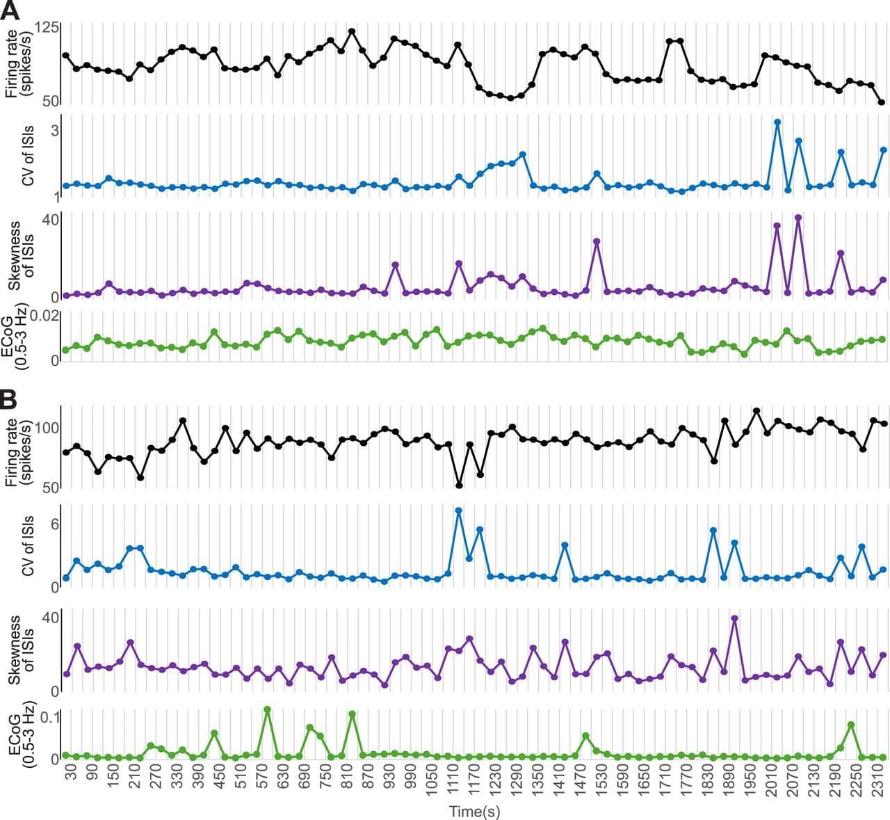
The first report of in vivo extracellular recordings in the external segment of the globus pallidus (GPe) of awake monkeys described that most GPe neurons show high-frequency spiking activity, interspersed with pauses (high-frequency discharge with pauses, HFD-P), while a smaller proportion was said to show low-frequency discharge with bursts (LFD-B; DeLong, 1971). Similar patterns of pallidal discharge have been demonstrated by other authors, both in primates (Katabi et al., 2022) and rodents (Bugaysen et al., 2010; Benhamou et al., 2012).
There is evidence, however, that the HFD-P and LFD-B subtypes of GPe neurons are only the most recognizable extremes of a continuous spectrum of properties of GPe neurons. This view is supported by in vivo and in vitro recordings in rodents which found that the firing properties of the population of GPe neurons distribute along a continuum, with specific cells firing within more limited boundaries of firing rates and patterns (Abdi et al., 2015; Cui et al., 2021). Furthermore, observations in rodents showed that GPe neurons display a wide range of firing rates and patterns (Deister et al., 2013). The firing pattern heterogeneity in in vivo recordings may arise, at least in part, due to shifts in firing behavior of the same neurons, as has been reported in rodent studies (Deister et al., 2013). Such variations in firing patterns may only be detectable when recordings extend over long time periods (Elias et al., 2008).
The loss of nigrostriatal dopamine fibers associated with parkinsonism induces multiple alterations in GPe, where neuronal firing becomes slower, may show more frequent bursts, and becomes more synchronized (Galvan et al., 2015; Courtney et al., 2023). However, the stability of firing patterns of GPe neurons in the parkinsonian state has not yet been investigated.
This talk is part of the “New Ideas in NCC Research” workshop of the Bamberg Mathematical Consciousness Science Initiative (BAMΞ). For more talks and details, see https://www.uni-bamberg.de/en/bamxi/r…
Abstract: In recent years, the search for the neural correlates of consciousness (NCC) has been complemented, and influenced, by the ongoing efforts to test neuroscientific theories of consciousness. A key insight from these efforts, though, is that many theories remain underdeveloped and not fully specified, making it harder to establish stringent tests for their predictions. In this talk, I will present a novel methodological approach that represents scientific theories as networks of beliefs structured in a core-periphery manner. These Prediction Maps visualize theoretical claims and empirical predictions, and illustrate their inferential relations. This framework further facilitates systematic theory testing by allowing researchers to evaluate the evidential weight of different components of a theory, and to identify which experimental results would constitute the most informative tests. To do so, we apply graph-theoretic and network analysis metrics, quantifying the centrality of specific predictions. I argue that this approach can advance efforts to arbitrate between theories of consciousness and to identify their most promising candidate mechanisms as NCCs.
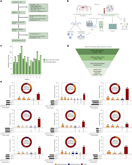
A personalized vaccine to treat glioblastoma, a fast-growing and incurable brain cancer that affects four in 100,000 people in the U.S., is safe and elicits robust and broad immune responses that appears to increase recurrence-free survival in a subset of patients after surgery, according to an early-stage clinical trial co-led by researchers at Washington University School of Medicine in St. Louis.
In patients with an especially aggressive form of glioblastoma, the vaccine caused no serious side effects and prolonged patients’ overall survival compared to historical outcomes after standard-of-care surgery and chemo-radiotherapy. One long-term survivor remains recurrence-free nearly five years later.
The results of the phase 1 trial, conducted at Siteman Cancer Center, based at Barnes-Jewish Hospital and WashU Medicine, were published May 12 in Nature Cancer. The study was led jointly by Mass General Brigham and Geneos Therapeutics, a Philadelphia-based biotechnology company.
“We are extremely encouraged by these results,” said Tanner M. Johanns, MD, PhD, lead author of the study and an assistant professor in the Division of Oncology in the John T. Milliken Department of Medicine at WashU Medicine. “This kind of vaccine is a first for glioblastoma, and it is exciting to think how we can leverage this individualized therapeutic DNA cancer vaccine platform to make a positive impact on the lives of patients who are fighting this disease. Additionally, combination therapies leveraging this personalized platform are currently being investigated at WashU to test if outcomes may be improved further.”
Abstract: Nature Cancer
Johanns and colleagues report the results (including safety, efficacy and immunogenicity) of a phase 1 clinical trial of a DNA-based personalized therapeutic cancer vaccine administered following surgical resection and radiation in patients with MGMT unmethylated glioblastoma.
When facing new situations or problems, humans typically rely on knowledge they acquired in the past. Specifically, neuroscience studies suggest that the brain reorganizes past experiences and previously acquired knowledge, creating mental frameworks that can help humans to solve the problems they are facing. The recombination of past knowledge into new mental structures also allows humans to flexibly plan future actions in changing environments. Past studies suggest that two key brain regions contribute to this process, the hippocampus and the medial prefrontal cortex (mPFC).
The hippocampus is a brain structure that plays a key role in the formation of memories and spatial navigation. The mPFC, on the other hand, is known to support decision-making, planning, reasoning and the integration of information.
Researchers at Beijing Normal University, the Chinese Academy of Medical Sciences, University College London (UCL) and other institutes recently set out to investigate how the hippocampus and mPFC work together to combine past knowledge into new configurations. Their findings, published in Nature Neuroscience, suggest that this process is supported by brief bursts of high-frequency neural activity in the hippocampus, called hippocampal ripples, and the replay (i.e., re-activation) of past experiences in the brain.