Researchers studied <i>LRRK2 </i> mutations in Parkinson’s disease. The mutations disrupted dopamine release in vulnerable neurons prior to neuronal loss, identifying potential targets and timing for intervention.

Researchers studied <i>LRRK2 </i> mutations in Parkinson’s disease. The mutations disrupted dopamine release in vulnerable neurons prior to neuronal loss, identifying potential targets and timing for intervention.

Thousands of skeletal human remains have been discovered with their brain tissue unexpectedly intact. Now, a team of scientists may have finally figured out why.

Modern therapies have transformed care for people with multiple sclerosis (MS). Yet the disease continues to progress in a substantial proportion of patients, even in the absence of relapses. Detecting and treating this gradual worsening of disability remains one of the greatest challenges in MS care. Researchers from the University of Basel, Switzerland, now report on a blood marker that could help track disease progression and monitor treatment response.
Relapses are only part of the disease process in MS. In some patients, physical and cognitive abilities continue to gradually decline even in the absence of relapses. Experts refer to this as progression independent of relapse activity (PIRA). This process develops slowly and is difficult to detect and quantify. A currently available blood test measures neurofilament light chain (NfL), a biomarker that mainly reflects neuroaxonal damage caused by acute inflammatory disease activity. A blood marker that captures the progressive component of the disease has so far been lacking.
“One of the key questions for us was whether progression leaves a different biological fingerprint than inflammation,” says Professor Jens Kuhle from the University of Basel and the Research Center for Clinical Neuroimmunology and Neuroscience (RC2NB) at University Hospital Basel. “If these processes are biologically distinct, we also need biomarkers that reflect different aspects of the disease rather than expecting a single marker to capture everything.”
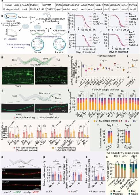
During context-dependent decision-making, anterior lateral motor cortex is required to configure stimulus-response mappings. Two-photon imaging reveals neurons that jointly encode context and choice, suggesting that flexible behavior arises from dynamic reconfiguration of local circuits rather than abstract context representations.
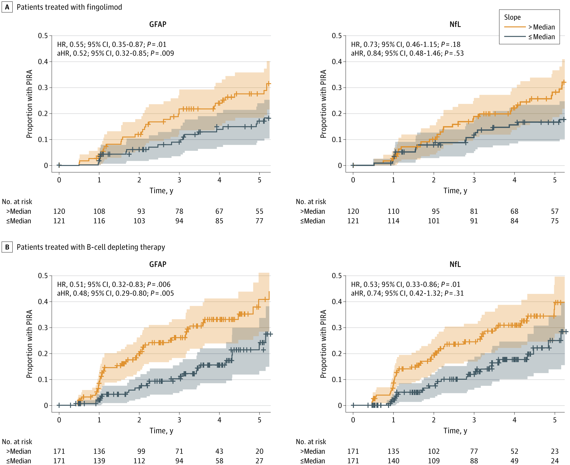
This cohort study evaluates the association between serum glial fibrillary acidic protein levels and future progression of multiple sclerosis.

Alzheimer’s disease is the most common form of dementia, which affects approximately 57 million people globally – a number that’s projected to rise to more than 150 million by 2050.
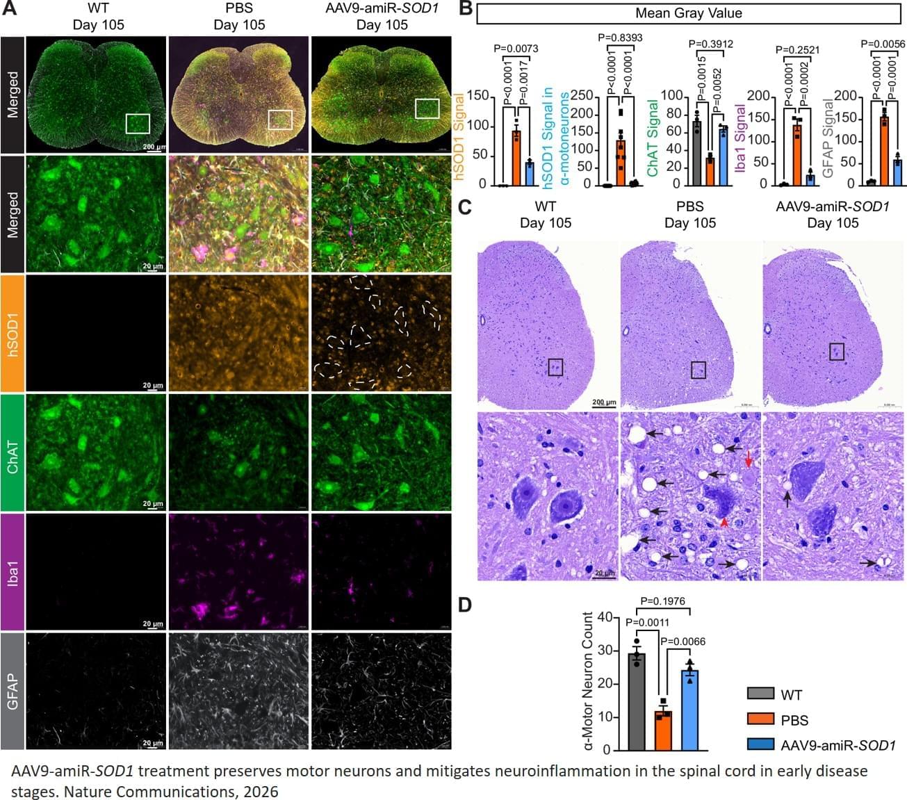
A single IV injection of a microRNA-based biologic suppressed production of the mutant SOD1 protein that causes amyotrophic lateral sclerosis (ALS); delayed disease onset by 60 days; and extended lifespan by 100 days, more than triple the average survival time, in mice models of the disease.
The gene therapy, delivered via adeno-associated virus (AAV) vector, preserved motor neurons and maintained neuromuscular connections in treated animals, which translated into improved muscle and respiratory function, motor performance, and lifespan in pre-clinical studies. These findings, published in Nature Communications, have the potential for clinical application in patients with SOD1-caused ALS, as well as other neurodegenerative diseases caused by toxic, gain-of-function gene mutations.
“These therapeutic benefits, from a single IV injection, are unprecedented among gene therapy approaches in this mouse model,” said the senior author. “No other studies have been able to achieve this kind of survival extension. This makes us very optimistic that our approach could have a meaningful impact for patients suffering from this horrible disease and warrants further clinical evaluation.”

Cerebral edema is the abnormal accumulation of fluid in any of the tissue compartments of the cerebral parenchyma. It remains a significant challenge in neurotrauma care because it contributes to secondary brain injury, affecting prognosis. This review analyzes the recent literature, including foundational studies, to describe the mechanisms of distinct types of cerebral edema following traumatic brain injury (TBI). Emerging concepts, such as the role of the glymphatic system and heme-derived inflammasomes, offer new insights into new types of edemas, differentiated by pathogenesis and potential treatments. Recent advancements in understanding these molecular mechanisms can improve therapeutic strategies, facilitating a better approach in the era of precision and personalized medicine.

Brain metastases are a major clinical concern in metastatic breast cancer, particularly in HER2-positive and triple-negative disease. Over time, approximately 25% to 50% of patients with these subtypes develop brain metastases, which can lead to neurological disability, cognitive impairment, reduced independence, and substantial deterioration in quality of life.
Despite this risk, routine brain imaging is not consistently recommended for patients without neurological symptoms.
An article published in The Breast argues that it may be time to reconsider this symptom-directed approach. Sarah Sammons, Nayan Lamba, and Nancy U. Lin outline a growing case for brain MRI screening in selected patients with metastatic breast cancer, while also acknowledging that survival benefit, optimal timing, cost-effectiveness, and potential harms remain uncertain.