A research team has identified a previously unknown enzyme, SIRT2, that plays a key role in memory loss associated with Alzheimer’s disease (AD). The study provides critical insights into how astrocytes contribute to cognitive decline by producing excessive amounts of the inhibitory neurotransmitter GABA.
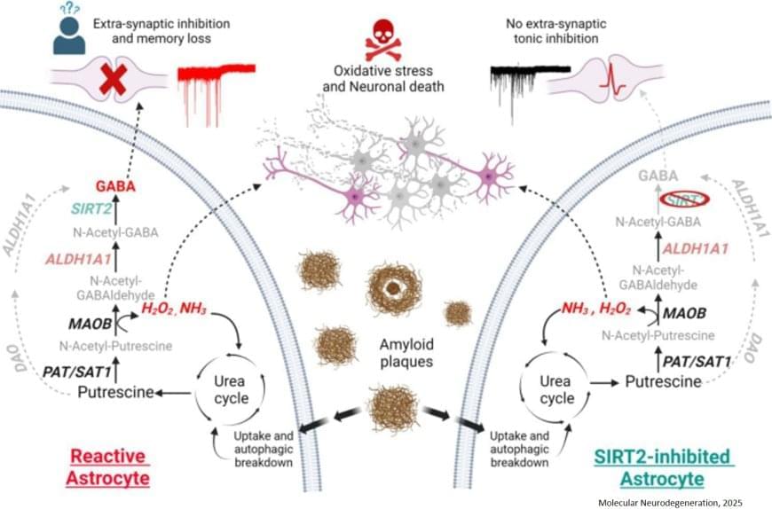
Astrocytes, once thought to only support neurons, are now known to actively influence brain function. In Alzheimer’s disease, astrocytes become reactive, meaning they change their behavior in response to the presence of amyloid-beta (Aβ) plaques, a hallmark of the disease. While astrocytes attempt to clear these plaques, this process triggers a harmful chain reaction. First, they uptake them via autophagy and degrade them by the urea cycle, as discovered in previous research. However, this breakdown results in the overproduction of GABA, which dampens brain activity and leads to memory impairment. Additionally, this pathway generates hydrogen peroxide (H2O2), a toxic byproduct that causes further neuronal death and neurodegeneration.
The research team set out to uncover which enzymes were responsible for excessive GABA production, hoping to find a way to selectively block its harmful effects without interfering with other brain functions. Using molecular analysis, microscopic imaging, and electrophysiology, the researchers identified SIRT2 and ALDH1A1 as critical enzymes involved in GABA overproduction in Alzheimer’s-affected astrocytes.