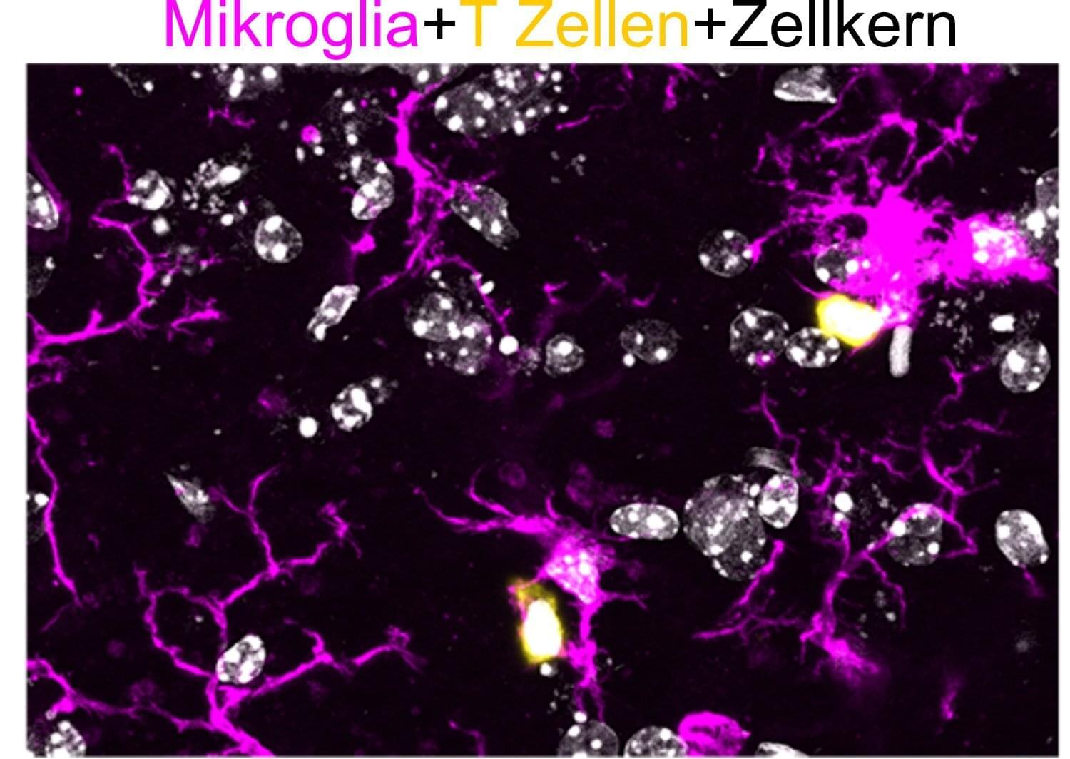
Toggle light / dark theme

Study identifies a key protein for healthy aging

Life expectancy and healthy aging in mice can be determined by a protein present in some cells of the immune system, according to a study published in the journal Cell Reports. When this protein—known as the CD300f immune receptor—is absent, animal models have a shorter life expectancy and suffer from pathologies associated with cognitive decline and premature aging, especially in females.

“Our study indicates that alterations in , for instance, in macrophages and microglia, can determine the healthy aging degree in mice,” notes Hugo Peluffo, leader of this study and member of the Faculty of Medicine and Health Sciences and the Institute of Neurosciences (UBneuro) of the University of Barcelona.

Understanding how the CD300f —and the myeloid cells of the immune system—can determine by themselves the onset rate of aging-associated pathologies, “will help to better understand this process, and it will contribute to the design of strategies to regulate its action. For instance, using the immune receptor CD300f as a target in biomedicine,” notes the expert. “Also, our team has previously shown that some variants of the CD300f immune receptor could be useful as biomarkers in patients.”

Ingestible Electronic Device Detects Breathing Depression in Patients

A new ingestible capsule can monitor vital signs from within the patient’s GI tract. The sensor could be used for less intrusive monitoring of sleep disorders such as sleep apnea, or for detecting opioid overdoses.

Diagnosing sleep disorders such as sleep apnea usually requires a patient to spend the night in a sleep lab, hooked up to a variety of sensors and monitors. Researchers from MIT, Celero Systems, and West Virginia University hope to make that process less intrusive, using an ingestible capsule they developed that can monitor vital signs from within the patient’s GI tract.

The capsule, which is about the size of a multivitamin, uses an accelerometer to measure the patient’s breathing rate and heart rate. In addition to diagnosing sleep apnea, the device could also be useful for detecting opioid overdoses in people at high risk, the researchers say.

Structure-related intrinsic electrical states and firing patterns of neurons with active dendrites

Activity of neurons embedded in networks is an inseparable composition of evoked and intrinsic processes. Prevalence of either component depends on the neuron’s function and state (e.g. low/high conductance or depolarization states). Dominant intrinsic firing is thought functionally normal for the pacemaker neuron, but not for the sensory afferent neuron or spinal motoneuron serving to transmit rather than to originate signals. Activity of the multi-functional networked cell, depending on its intrinsic states, bears both cell-and network-defined features. Complex firing patterns of a neuron are conventionally attributed to complex spatial-temporal organization of inputs received from the network-mates via synapses, in vast majority dendritic. This attribution reflects widespread views of the within-cell job sharing, such that the main function of the dendrites is to receive signals and deliver them to the axo-somatic trigger zone, which actually generates the output pattern. However, these views require revisiting with account of active properties of the dendrites due to voltage-dependent channels found in the dendritic membrane of practically all types of explored neurons. Like soma and axon, the dendrites with active membrane are able to generate self-maintained, propagating depolarizations and thus share intrinsic pattern-forming role with the trigger zone. Unlike the trigger zone, the dendrites have complex geometry, which is subject to developmental, activity-dependent, or neurodegenerative changes. Structural features of the arborization inevitably impact on electrical states and cooperative behavior of its constituting parts at different levels of organization, from branches and sub-trees to voltage-and ligand-gated ion channels populating the membrane. Nearly two decades of studies have brought numerous phenomenological demonstrations of influence of the dendritic structure on firing patterns in neurons. A necessary step forward is to comprehend these findings and build a firm theoretical basis, including quantitative relationships between geometrical and electrical characteristics determining intrinsic firing of neurons. This Research Topic is aimed at bringing together contributions of researches from different domains of expertise and building a conceptual framework for deeper insight into the nature of dynamic intrinsic motifs in the firing patterns.

We welcome research and methodology papers, mini-reviews, conceptual generalizations and opinions on the following issues:

1. Electrical states of heterogeneous populations of ion channels: definition, life-times, meta-and multi-stability.

8: Spike Trains — Intro to Neural Computation

MIT 9.40 Introduction to Neural Computation, Spring 2018
Instructor: Michale Fee.
View the complete course: https://ocw.mit.edu/9-40S18
YouTube Playlist: https://www.youtube.com/playlist?list=PLUl4u3cNGP61I4aI5T6OaFfRK2gihjiMm.

Covers extracellular spike waveforms, local field potentials, spike signals, threshold crossing, the peri-stimulus time histogram, and the firing rate of a neuron.

License: Creative Commons BY-NC-SA
More information at https://ocw.mit.edu/terms.
More courses at https://ocw.mit.edu.

We encourage constructive comments and discussion on OCW’s YouTube and other social media channels. Personal attacks, hate speech, trolling, and inappropriate comments are not allowed and may be removed.

More details at https://ocw.mit.edu/comments

6: Dendrites — Intro to Neural Computation

MIT 9.40 Introduction to Neural Computation, Spring 2018
Instructor: Michale Fee.
View the complete course: https://ocw.mit.edu/9-40S18
YouTube Playlist: https://www.youtube.com/playlist?list=PLUl4u3cNGP61I4aI5T6OaFfRK2gihjiMm.

Covers the dendrite circuit diagram, voltage plot, length diagram, dendritic radius, electronic length, and the circuit diagram of a two-compartment model.

License: Creative Commons BY-NC-SA
More information at https://ocw.mit.edu/terms.
More courses at https://ocw.mit.edu.

We encourage constructive comments and discussion on OCW’s YouTube and other social media channels. Personal attacks, hate speech, trolling, and inappropriate comments are not allowed and may be removed.

More details at https://ocw.mit.edu/comments

Study unveils the engagement of different cortical networks while humans are unconscious

Despite substantial work, we are still unsure which brain regions are involved and how they are impacted when consciousness is disrupted.


States of unconsciousness, such as those that occur during sleep or while under the effect of anesthesia, have been the focus of countless past neuroscience studies. While these works have identified some brain regions that are active and inactive when humans are unconscious, the precise contribution of each of these regions to consciousness remains largely unclear.

Researchers at Massachusetts General Hospital recently carried out a study aimed at better understanding the activity of different regions of the cortex, the outer layer of the mammalian , during different states of unconsciousness, namely sleep and . Their paper, published in Neuron, identifies distinct cortical networks that are engaged during different states of unconsciousness.

“We have always been interested in trying to understand better how in the brain gives rise to consciousness,” Dr. Rina Zelmann, the lead researcher for the study, told Medical Xpress. “This is a huge and difficult question to answer. In this project, we started with seemingly simple questions, such as: What happens in the human brain when we are unconscious? And, what happens when we cannot be awakened?”