A nasal spray using extracellular vesicles can reduce brain inflammation, restore cell function, and improve cognition, offering a potential breakthrough in reversing brain aging.
Scientists from Johns Hopkins Medicine report new evidence that clusters of brain tissue derived from the cells of patients with Alzheimer’s disease may be used to evaluate how certain patients with the neurodegenerative condition may respond to drugs commonly prescribed to treat psychiatric symptoms of the disorder. The findings, based on a study of lab-grown brain tissues known as organoids, contribute to a growing body of evidence that brain organoids may also one day be used to more precisely develop and prescribe treatments for subgroups of patients with Alzheimer’s disease, which is the most common form of dementia, and affects more than seven million Americans.
In addition, the researchers found that tiny particles, known as extracellular vesicles, which are secreted by organoids, may contain cellular information that could help scientists find new biomarkers to diagnose and stage Alzheimer’s disease. A report of the findings is published in Alzheimer’s & Dementia.
“Our study suggests that large-scale, patient-derived brain organoids and the vesicles they secrete can help us stage Alzheimer’s disease, investigate the mechanisms that drive it and assess how patient subgroups may respond to different treatments,” says study leader Vasiliki Machairaki, Ph.D., associate professor of genetic medicine at the Johns Hopkins University School of Medicine.
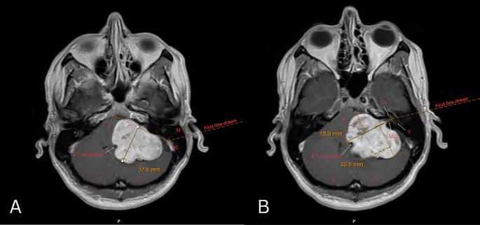
Horner’s syndrome (HS) occurs when there is interruption of the oculosympathetic pathway (OSP). This article reviews the anatomy of the OSP and clinical findings associated with lesions located at various positions along this pathway. The imaging findings of lesions associated with HS at various levels of the OSP, classified as preganglionic HS (first-and second-order neuron HS) or postganglionic HS (third-order neuron HS), are demonstrated.
The authors discovered that training mice to exhibit a placebo effect with one type of pain produces marked relief of several different types of pain, including pain caused by injury.
To establish that the native opioid peptides actually drive pain relief, similar to opioid painkillers such as morphine, the researchers employed a light-activated drug developed in Banghart’s lab called PhNX, for photoactivatable naloxone. Naloxone, also known as Narcan, is the medicine used to reverse opioid overdoses by blocking opioid receptors. Using light, they were able to precisely control the site and timing of opioid signaling interference. Using PhNX, the scienists found that both morphine-induced pain relief and placebo pain relief rely on opioid signaling in the vlPAG brain region.
Co-first author: “We essentially trained a mouse brain to create its own broad-spectrum painkillers on demand, precisely where they are needed to treat pain, without the off-target effects of opioid-based painkillers.”
“These results increase the translational relevance of rodent placebo models to clinical contexts, in which patients’ prior experiences with drugs and treatment settings can generalize to broader expectations of improvement,” the researchers conclude in their paper. ScienceMission sciencenewshighlights.
Placebo effects, in which patients experience relief without therapeutic treatment, increasingly have been considered as potentially powerful clinical treatments for ailments such as depression and pain. Yet the neurological mechanisms underlying such processes are not fully understood.
Now, a multi-institutional team has pinpointed the brain circuitry responsible for placebo pain relief. Their findings, reported in the journal Neuron, describe brain regions that support placebo effects and identify sites where endogenous opioid neuropeptides (commonly referred to as endorphins) provide signals that are critical for placebo pain relief.
This is one of my favourite comparisons: polymathy is cognitive biodiversity.
Monoculture farming depletes soil, invites disease, collapses under pressure. One blight, one drought and the whole field dies.
Why do we accept the same fragility in how we think?
The specialist mind is similar to a monoculture. Trained to the depth in one domain and optimized for known conditions. When the paradigm breaks, it can only do what it has always done.
Functional unblinding was common in most psychedelic randomized clinical trials for psychiatric disorders, with 70% correctly identifying treatment allocation, raising concerns for trial validity.
Question What is the prevalence of blinding integrity assessment and the extent of functional unblinding in psychedelic randomized clinical trials (RCTs) for psychiatric disorders?
Findings Of 112 RCTs identified, 29.5% (n = 33) evaluated blinding integrity. Functional unblinding was substantial: psilocybin, lysergic acid diethylamide (LSD), and ayahuasca studies frequently reported blinding failure values of more than 90% among participants and raters; inert placebo-controlled 3,4-methylenedioxymethamphetamine (MDMA) trials exceeded 85%; ketamine trials rarely assessed blinding (17.9%) but showed improved preservation with midazolam vs saline controls.
Meaning Functional unblinding is pervasive in psychedelic RCTs, underscoring the need for standardized assessment methods and improved trial designs to ensure valid efficacy evaluations.
Despite its small size—typically only several inches, beak to tail—the zebra finch is a remarkable learner. A songbird native to Australia, it’s renowned for its ability to pick up new songs.
That talent has made it a favorite of scientists studying how animal brains imprint new skills, particularly vocal learning, or the capacity to perfect new sounds. And now researchers at Boston University have discovered another quirk to the zebra finch brain—one that could also have implications for understanding our own gray matter.
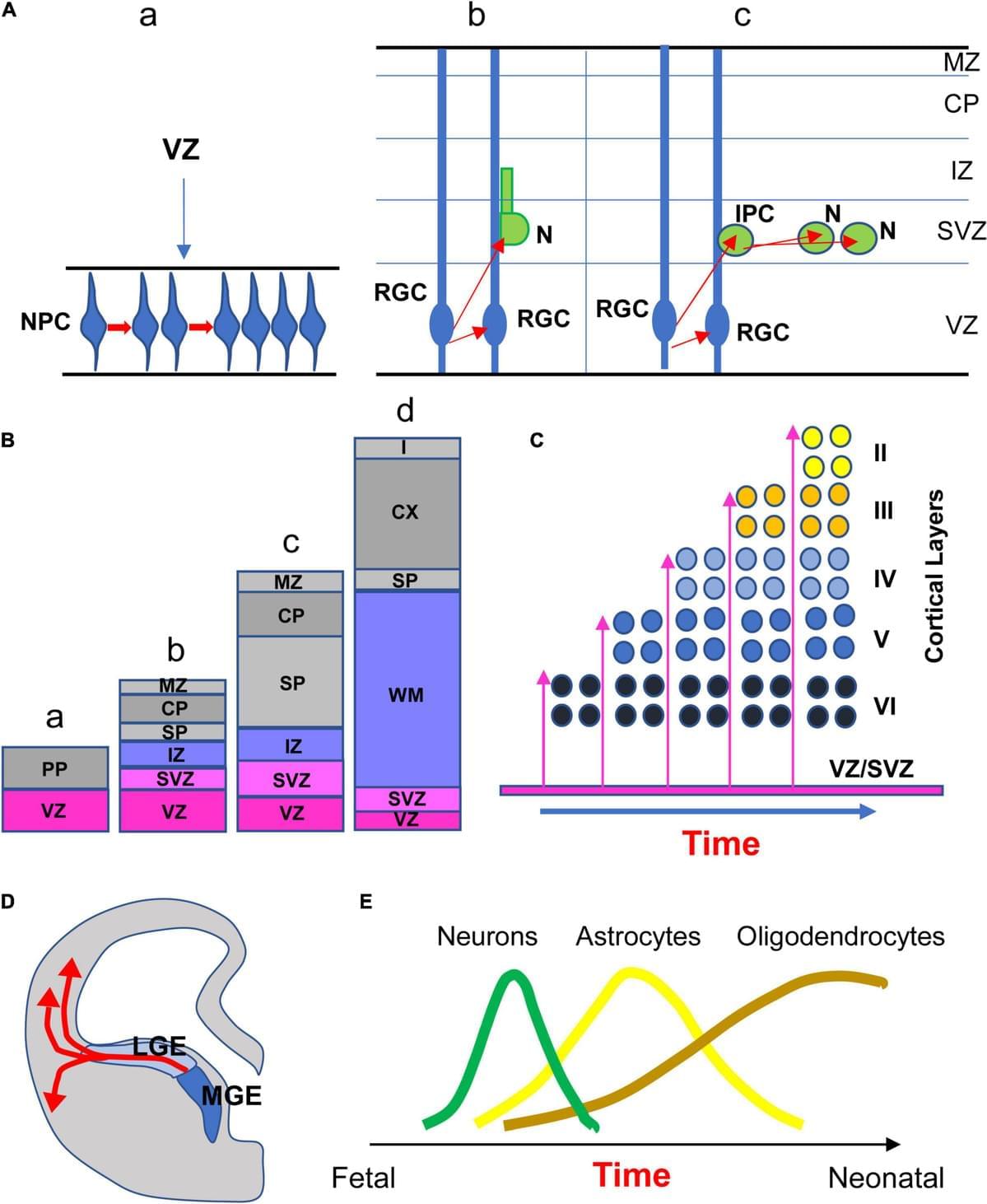
In a study that looked at the bird’s brain in unprecedented detail, they uncovered new insights into a mechanism known as neurogenesis—the birth, migration, and maturation of neurons—that may help the brain learn, add new skills, and restore and repair itself.
Observing the finch brain using a high-powered microscope, the researchers watched as new neurons bullied their way through the brain en route to bolstering existing circuits and connections. They’d expected the neurons to gingerly step around established brain structures, including more mature brain cells, to better preserve them; instead, they saw the neurons tunnel right through, squishing and shoving as they went. That may help the birds learn new things or repair damage, but it could also come with a cost to existing cells and memories.
According to the BU-led team, their findings could help explain why neurogenesis may not occur in humans beyond the womb, increasing our vulnerability to a range of brain disorders. The findings were published in Current Biology.
Abstract: Current Biology
Read More
Following out-of-hospital cardiac arrest, targeted hypothermia did not affect societal participation or cognitive function at 24 months compared with normothermia; most recovery occurred within 6 months.
Question Does hypothermia after out-of-hospital cardiac arrest affect societal participation or cognitive functioning at 24 months post arrest, and how do these outcomes evolve over time?
Findings This follow-up of the randomized clinical Targeted Hypothermia vs Targeted Normothermia After Out-of-Hospital Cardiac Arrest trial found no significant differences in societal participation or cognitive functioning between targeted hypothermia and normothermia at 24 months. Overall recovery was limited beyond 6 months.
Meaning Targeted hypothermia compared with normothermia did not affect outcomes 24 months post arrest, suggesting no longer-term effect of hypothermia for the explored outcomes; 6 months may suffice as an end point when assessing functional or cognitive outcomes after out-of-hospital cardiac arrest.