Toggle light / dark theme

Study Reveals Critical Age When Your Thinking Begins to Decline

As Shakespeare put it, we all have our entrances and our exits on this grand stage we call life, and now researchers have identified the specific point in middle-age when our brain cells show the first signs of starting down a downward slope.

That age, based on brain scans and tests covering 19,300 individuals, is on average around 44 years. It’s here that degeneration starts to be noticeable, before hitting its most rapid rate at age 67. By the time we reach 90, the speed of brain aging levels off.

According to the team behind the new study, led by researchers from Stony Brook University in the US, the findings could be helpful in figuring out ways to promote better brain health during the later stages of life.

A torpor-like state in mice slows blood epigenetic aging and prolongs healthspan

Dissecting the effects of hypothermic and hypometabolic states on aging processes, the authors show that activation of neurons in the preoptic area induces a torpor-like state in mice that slows epigenetic aging and improves healthspan. These pro-longevity effects are mediated by reduced Tb, reinforcing evidence that Tb is a key mediator of aging processes.

Rosemary Compound Shows Promise for Alzheimer’s Disease

Researchers developed a stable form of carnosic acid, a compound found in rosemary and sage. The compound showed promise for improving learning and memory and reducing signs of Alzheimer’s disease in mouse models. The corresponding study was published in Antioxidants. The researchers hope to test the compound in human trials.

Carnosic acid has both antioxidant and anti-inflammatory properties. While pure carnosic acid is too unstable for medical use, the new compound developed by scientists from Scripps Research fully converts to carnosic acid in the gut before absorption into the bloodstream. They called the compound ‘diAcCA’

As a part of the study, the researchers tested the compound’s efficacy in mouse models for three months. Ultimately, the compound improved learning and memory, and reduced markers of inflammation, formation of amyloid plaques, and phosphorylated tau aggregates.

Excitation-Inhibition Balance Controls Information Encoding in Neural Populations

Understanding how the complex connectivity structure of the brain shapes its information-processing capabilities is a long-standing question. By focusing on a paradigmatic architecture, we study how the neural activity of excitatory and inhibitory populations encodes information on external signals. We show that at long times information is maximized at the edge of stability, where inhibition balances excitation, both in linear and nonlinear regimes. In the presence of multiple external signals, this maximum corresponds to the entropy of the input dynamics. By analyzing the case of a prolonged stimulus, we find that stronger inhibition is instead needed to maximize the instantaneous sensitivity, revealing an intrinsic tradeoff between short-time responses and long-time accuracy.

Why zero could unlock how the brain perceives absence

When I’m birdwatching, I have a particular experience all too frequently. Fellow birders will point to the tree canopy and ask if I can see a bird hidden among the leaves. I scan the treetops with binoculars but, to everyone’s annoyance, I see only the absence of a bird.

Our mental worlds are lively with such experiences of absence, yet it’s a mystery how the mind performs the trick of seeing nothing. How can the brain perceive something when there is no something to perceive?

For a neuroscientist interested in consciousness, this is an alluring question. Studying the neural basis of ‘nothing’ does, however, pose obvious challenges. Fortunately, there are other – more tangible – kinds of absences that help us get a handle on the hazy issue of nothingness in the brain. That’s why I spent much of my PhD studying how we perceive the number zero.

New 4D Brain Map reveals potential early warning signs of multiple sclerosis

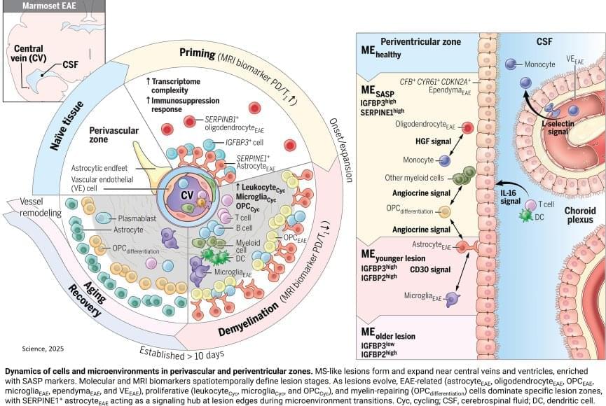
To mimic the conditions of the human brain, the researchers opted not to use a mouse model for MS, instead advancing a model that uses the marmoset, a nonhuman primate. Compared to mouse brains, marmoset and human brains have a higher ratio of white matter (the “wires” of the brain) to gray matter (neuronal cell bodies). The marmoset model creates multiple lesions that closely resemble those seen in human MS and that can be tracked in real time using MRI imaging. Because these lesions can be induced experimentally, the model offers a look at the earliest stages of inflammation and immune responses that lead to MS-like demyelination.

One key player identified was a specific type of astrocyte, one of the support cell types in the brain, that turns on a gene called SERPINE1 or plasminogen activator inhibitor-1 (PAI1). They found SERPINE1-expressing astrocytes in vulnerable brain borders before visible damage occurs, clustering near blood vessels and the fluid-filled ventricles of the brain and signaling future areas of lesion development. These astrocytes also appeared to influence the behavior of other cells near the lesion area, including the ability of immune cells to enter the brain and contribute to inflammation, as well as the precursor cells involved in myelin repair.

Given that SERPINE1-expressing astrocytes accumulated at the edges of growing lesions, where damage happens but healing also begins, their potential dual role in coordinating signals that could lead to either tissue repair or further damage was an unexpected wrinkle that will require further study. It’s possible that the earliest responses could be a part of a protective mechanism that becomes overwhelmed as the injury progresses. It’s also possible that the same mechanism could itself become disease-causing.


Using an animal model of multiple sclerosis (MS), researchers have created a four-dimensional brain map that reveals how lesions similar to those seen in human MS form. These findings, published in Science, provide a window into the early disease state and could help identify potential targets for MS treatments and brain tissue repair.

The researchers combined repeated MRI imaging with brain-tissue analysis, including gene expression, to track the onset and development of MS-like lesions. They uncovered a new MRI signature that can help detect brain regions at risk for damage weeks before any visible lesions occur. They also identified “microenvironments” within affected brain tissue based on observed patterns of neural function, inflammation, immune and support cell responses, gene expression, and levels of damage and repair.

“Identifying the early events that occur after inflammation and teasing apart which are reparative versus which are damaging, can potentially help us identify MS disease activity sooner and develop treatments to slow or stop its progression,” said the author.

No evidence for ‘wind turbine syndrome’ claims: Windmill noise is no more stressful than traffic sounds, study suggests

A team of cognitive neuroscientists and acoustic engineers at Adam Mickiewicz University, in Poland, has found no evidence that wind turbine noise causes mental impairment. In their study, published in the journal Humanities and Social Sciences Communication, the group conducted experiments exposing human volunteers to various noises and measured a range of impacts.

Over the past several years, several groups and individuals around the world, most particularly in the U.S., have conceived of the idea of something called “wind turbine syndrome”—a theory that suggests noise from windmills can cause , or other health problems such as cancer. To date, such claims have not been backed up by research or any other type of proof. In this new effort, the research team in Poland sought to find out if there is any merit to the theory.

The researchers recruited 45 students at a local university who listened to various noises while wearing devices that measured their brainwaves. The researchers intentionally chose young volunteers because prior research has shown they are more sensitive to noise than .

Scientists Have Discovered Shocking Amounts of Microplastics in the Brain — And It Could Be Increasing Our Risk of Dementia

The brain has higher concentrations of plastic particles compared to other organs, with increased levels found in dementia patients.

In a comprehensive commentary published in Brain Medicine, researchers highlight alarming new evidence of microplastic accumulation in human brain tissue, offering critical insights into potential health implications and prevention strategies. This commentary examines findings from a groundbreaking Nature Medicine article by Nihart et al. (2025) on the bioaccumulation of microplastics in the brains of deceased individuals.

The research reveals that human brains contain approximately a spoonful of microplastics and nanoplastics (MNPs), with levels three to five times higher in individuals with documented dementia diagnoses. Even more concerning, brain tissue exhibited MNP concentrations seven to thirty times higher than those found in other organs, such as the liver or kidneys.

Optimal brain processing requires balance between excitatory and inhibitory neurons, study suggests

The brain’s ability to process information is known to be supported by intricate connections between different neuron populations. A key objective of neuroscience research has been to delineate the processes via which these connections influence information processing.

Researchers at the University of Padova, the Max Planck Institute for the Physics of Complex Systems and École Polytechnique Fédérale de Lausanne recently carried out a study aimed at better understanding the contribution of excitatory and inhibitory neuron populations to the brain’s encoding of information. Their findings, published in Physical Review Letters, show that is maximized when the activity of excitatory and inhibitory neurons is balanced.

“Our research was inspired by a fundamental question in neuroscience: how does the structure of the brain shape its ability to process information?” Giacomo Barzon, co-author of the paper, told Medical Xpress. “The brain continuously receives and integrates sensory inputs, and neurons do not act in isolation—they are part of complex, recurrent networks. One particularly intriguing feature of these networks is the balance between the activity of excitatory and inhibitory neurons, which has been observed across different brain regions.”