Toggle light / dark theme

Scientists Have Discovered Shocking Amounts of Microplastics in the Brain — And It Could Be Increasing Our Risk of Dementia

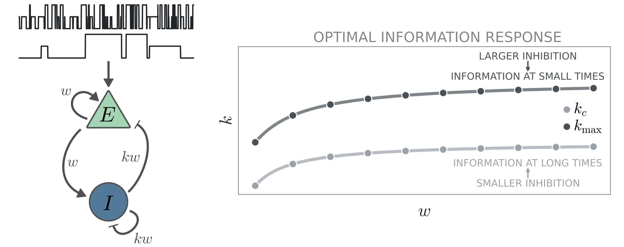
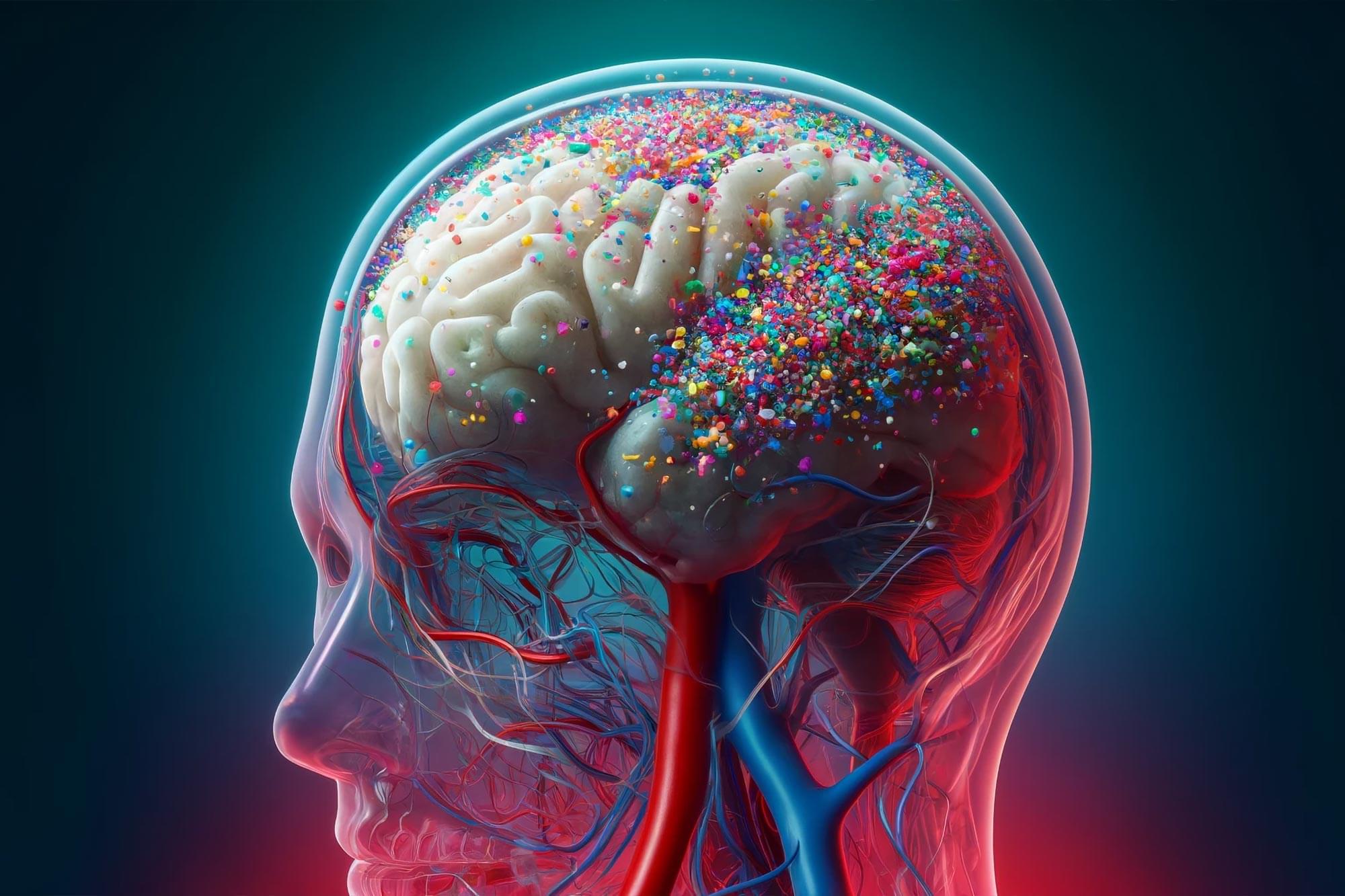
The brain has higher concentrations of plastic particles compared to other organs, with increased levels found in dementia patients.

In a comprehensive commentary published in Brain Medicine, researchers highlight alarming new evidence of microplastic accumulation in human brain tissue, offering critical insights into potential health implications and prevention strategies. This commentary examines findings from a groundbreaking Nature Medicine article by Nihart et al. (2025) on the bioaccumulation of microplastics in the brains of deceased individuals.

The research reveals that human brains contain approximately a spoonful of microplastics and nanoplastics (MNPs), with levels three to five times higher in individuals with documented dementia diagnoses. Even more concerning, brain tissue exhibited MNP concentrations seven to thirty times higher than those found in other organs, such as the liver or kidneys.

Optimal brain processing requires balance between excitatory and inhibitory neurons, study suggests

The brain’s ability to process information is known to be supported by intricate connections between different neuron populations. A key objective of neuroscience research has been to delineate the processes via which these connections influence information processing.

Researchers at the University of Padova, the Max Planck Institute for the Physics of Complex Systems and École Polytechnique Fédérale de Lausanne recently carried out a study aimed at better understanding the contribution of excitatory and inhibitory neuron populations to the brain’s encoding of information. Their findings, published in Physical Review Letters, show that is maximized when the activity of excitatory and inhibitory neurons is balanced.

“Our research was inspired by a fundamental question in neuroscience: how does the structure of the brain shape its ability to process information?” Giacomo Barzon, co-author of the paper, told Medical Xpress. “The brain continuously receives and integrates sensory inputs, and neurons do not act in isolation—they are part of complex, recurrent networks. One particularly intriguing feature of these networks is the balance between the activity of excitatory and inhibitory neurons, which has been observed across different brain regions.”

Age and cognitive skills: Use it or lose it

Regular use of math and reading skills could prevent cognitive decline with age, according to a new Science Advances study.


Cognitive skills of the population such as literacy and numeracy are important not only for individual incomes but also for the economic growth of nations (26). As a result, the aging of world populations presents an economic concern if the commonly assumed declines of these skills with age hold.

We use longitudinal variation in individual literacy and numeracy skills for a representative adult sample to create age-skill profiles that credibly separate age from cohort effects. The pure age component that we derive provides a different perspective on the impacts of aging populations. Overall, our results are not consistent with a view that a natural law dictates an inevitable decline in these skills with age. Potential cognitive declines only occur at later ages and are not inevitable with usage of skills.

This is consolation for countries with aging populations, but avoidance of skill losses is not automatic and appears related to stimulation from skill usage. These results thus suggest that age-skill relationships of adults deserve policy attention, consistent with concerns about the necessity of lifelong learning.

AI companions and Mental health: Can Virtual Companions reduce Loneliness?

You may have noted that AI companions are an intriguing yet complex phenomenon in this modern age. This event has caused various psychological effects on the human mind, both beneficial and detrimental. With the rapid advancement of AI systems, including conversational interfaces, virtual personal assistants, and robotic companions, interactions with these technologies are increasingly influencing emotional well-being and social behavior. You must have watched the film “Her,” which highlights similar themes of companionship with an operating system.

Her follows Theodore, a lonely writer who develops a deep emotional connection with an AI assistant named Samantha. Unlike traditional AI, Samantha isn’t just a programmed voice—she learns, evolves, and expresses emotions, making Theodore feel truly seen and understood. Their relationship blurs the line between human and machine companionship, raising questions about whether AI can meet emotional needs the way real human relationships do. As Samantha grows beyond Theodore, the film explores what it means to love something that isn’t physically present and whether AI relationships can ever replace real human connection.

This article will explore the psychological effects of AI companions, their potential benefits, and the limitations of this technology.

Measurement of CSF flow and brain motion in Chiari malformation type I subjects undergoing posterior fossa decompression surgery

Radiologically, Chiari malformation type I (CM-I) is characterized by cerebellar tonsil herniation of at least 5 mm through the foramen magnum. In symptomatic cases, posterior fossa decompression (PFD) surgery is often performed and improves symptoms in approximately 75% of patients. However, the surgery involves risks, and identifying which candidates will benefit from surgery is important. It has previously been shown that the amount of tonsillar descent does not correlate with symptom severity or surgical outcomes. The authors hypothesized that using advanced neuroimaging methods to directly measure CSF flow and brain motion will give insights regarding which patients have the greatest likelihood of cerebral dynamic improvements from surgery.

Here, the authors evaluated 108 CM-I patients (age 19–70 years), 61 of whom underwent PFD surgery. The authors used phase-contrast MRI to measure CSF flow/stroke volume and cine displacement encoding with stimulated echoes (DENSE) imaging to measure brain motion, with a goal to predict postsurgical cerebral dynamic improvements from presurgical images.

The authors found that CSF stroke volume increased after PFD surgery by 28.9% (p = 0.014), brainstem motion decreased after surgery by 17.3% (p = 0.002), and cerebellum motion decreased 45.2% (p < 0.001). Notably, the amount of CSF flow increase after surgery had no relationship to tonsillar descent (R = 0.059, p = 0.767) but did relate to the amount of presurgical CSF flow (R = −0.518, p = 0.005). Likewise, improvements to brain motion were better predicted by the amount of presurgical motion (brainstem, R = −0.638, p < 0.001; cerebellum, R = −0.878, p < 0.001) than by tonsillar descent (brainstem, R = −0.312, p = 0.093; cerebellum, R = −0.620, p < 0.001).

Nasal spray to treat traumatic brain injury

A new study suggests a nasal spray developed to target neuroinflammation could one day be an effective treatment for traumatic brain injury (TBI). By studying the effects of the nasal anti-CD3 in a mouse model of TBI, researchers found the spray could reduce damage to the central nervous system and behavioral deficits, suggesting a potential therapeutic approach for TBI and other acute forms of brain injury. The results are published in Nature Neuroscience.

The study examines the monoclonal antibody Foralumab, made by Tiziana, which has been tested in clinical trials for patients with multiple sclerosis, Alzheimer’s disease, and other conditions.

Multiple experiments were done in mouse models with moderate-to-severe traumatic brain injury to explore the communication between regulatory cells induced by the nasal treatment and the microglial immune cells in the brain. Over time, researchers were able to identify how they modulate immune response.

In addition to assessing the effects of the treatment, the research team was able to learn about immune response over time and compare the immune responses and effects of TBI in the mice.

The next step in the research is to translate the findings from preclinical models to human patients.

Scientists Identify Key Protein Driving Alzheimer’s Brain Cell Death

Scientists have identified the 1N4R tau isoform as a key driver of Alzheimer’s.

Alzheimer’s disease is a progressive neurological disorder that primarily affects older adults, leading to memory loss, cognitive decline, and behavioral changes. It is the most common cause of dementia. The disease is characterized by the buildup of amyloid plaques and tau tangles in the brain, which disrupt cell function and communication. There is currently no cure, and treatments focus on managing symptoms and improving quality of life.

Exercise training increases size of hippocampus and improves memory

Deterioration of the hippocampus precedes and leads to memory impairment in late adulthood (1, 2). Strategies to fight hippocampal loss and protect against the development of memory impairment has become an important topic in recent years from both scientific and public health perspectives. Physical activity, such as aerobic exercise, has emerged as a promising low-cost treatment to improve neurocognitive function that is accessible to most adults and is not plagued by intolerable side effects often found with pharmaceutical treatments (3). Exercise enhances learning and improves retention, which is accompanied by increased cell proliferation and survival in the hippocampus of rodents (46); effects that are mediated, in part, by increased production and secretion of BDNF and its receptor tyrosine kinase trkB (7, 8).

Aerobic exercise training increases gray and white matter volume in the prefrontal cortex (9) of older adults and increases the functioning of key nodes in the executive control network (10, 11). Greater amounts of physical activity are associated with sparing of prefrontal and temporal brain regions over a 9-y period, which reduces the risk for cognitive impairment (12). Further, hippocampal and medial temporal lobe volumes are larger in higher-fit older adults (13, 14), and larger hippocampal volumes mediate improvements in spatial memory (13). Exercise training increases cerebral blood volume (15) and perfusion of the hippocampus (16), but the extent to which exercise can modify the size of the hippocampus in late adulthood remains unknown.

To evaluate whether exercise training increases the size of the hippocampus and improves spatial memory, we designed a single-blind, randomized controlled trial in which adults were randomly assigned to receive either moderate-intensity aerobic exercise 3D/wk or stretching and toning exercises that served as a control. We predicted that 1 y of moderate-intensity exercise would increase the size of the hippocampus and that change in hippocampal volume would be associated with increased serum BDNF and improved memory function.