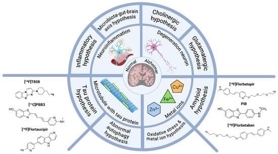
R&d and field deployment of orchestrated objective reduction

The metabolic fitness of microglia is markedly impaired in TREM2 knockout (KO) models [58]. TREM2, through its adaptors DAP12 and DAP10, activates the mechanistic target of rapamycin (mTOR) signaling pathway, which plays a crucial role in regulating metabolic pathways and protein synthesis [11, 58]. Loss of TREM2 impairs mTOR activation, leading to reduced ATP production and biosynthesis. In vivo FDG-PET imaging of TREM2 KO and TREM2 T66M knock-in mice shows a significant reduction in cerebral glucose metabolism [67, 68]. This decrease may correlate with impaired glucose uptake by microglia. Supporting this, ex vivo measurements of isolated microglia from TREM2 KO animals reveal lower FDG uptake [68].
Given the pivotal role of microglial metabolism in AD, targeting this process represents a promising therapeutic strategy. Agents such as interferon-γ (IFN-γ) and cyclocreatine, which enhance ATP production, have been shown to restore microglial functions and mitigate AD pathology [58, 65]. Notably, TREM2-activating antibodies boost microglial energy metabolism by promoting mitochondrial fatty acid and glucose oxidation [69]. Moreover, translocator protein (TSPO)-PET and FDG-PET imaging have demonstrated that TREM2 activation enhances microglial activity and glucose metabolism in amyloid mouse models. Thus, targeting TREM2 and microglial metabolism may complement existing AD therapies, which primarily focus on amyloid clearance and synaptic dysfunction, providing a more comprehensive approach to disease intervention.
Lipid metabolism is crucial for maintaining microglial functions and CNS homeostasis, influencing cellular membrane integrity, energy storage, and inflammatory responses. Emerging evidence identifies TREM2 as a key regulator of lipid metabolism in the brain. TREM2 binds a diverse range of lipids, including anionic and zwitterionic species such as sphingomyelin, phosphatidic acid, phosphatidylinositol, phosphatidylcholine, phosphatidylglycerol, phosphatidylserine (PtdSer) and sulfatide [49, 53, 70]. Among these, PtdSer is the most abundant negatively charged phospholipid in the inner leaflet of the plasma membrane in eukaryotic cells [71]. In neurodegenerative conditions, PtdSer externalization on damaged or apoptotic neurons serves as an “eat-me” signal, triggering TREM2-dependent microglial synaptic pruning and cell clearance [72]. Super-resolution microscopy and in vivo imaging studies have demonstrated that Aβ oligomer-induced hyperactive synapses expose PtdSer, marking them for TREM2-mediated engulfment, which helps mitigate neuronal hyperactivity in AD models. Additionally, individuals carrying TREM2 loss-of-function variants exhibit an accumulation of apoptotic-like synapses [72], underscoring TREM2’s essential role in synaptic homeostasis during early AD pathology. Beyond synaptic pruning, TREM2 facilitates the recognition and clearance of damaged cells. Notably, over-expression of TREM2 in non-phagocytic cells, such as Chinese hamster ovary (CHO) and HEK293 cells, enables them to engulf apoptotic neurons, highlighting TREM2’s function in lipid sensing and phagocytosis [16, 73]. This broad lipid-binding capability underscores TREM2’s critical role in modulating microglial responses to neurodegenerative insults and preserving neuronal health.
A new study from Columbia University Mailman School of Public Health and the Columbia Butler Aging Center suggests that risk factors and biomarkers related to Alzheimer’s disease are associated with cognition much earlier in life than previously recognized. The study highlights significant associations between cognition and Alzheimer’s disease risk factors as young as ages 24 to 44 and underscores the importance of early prevention.
This is the first study to systematically examine Alzheimer’s disease risk factors, including biomarkers related to cognitive impairment in a large group of generally healthy middle-aged individuals in the U.S. The findings are published in The Lancet-Regional Health Americas.
“Previously, research on Alzheimer’s disease risk factors has focused on individuals aged 50 and older,” said Allison Aiello, Ph.D., James S. Jackson Healthy Longevity Professor of Epidemiology in the Butler Aging Center and Columbia Mailman School. “The potential impact of our findings is substantial, offering clinicians and health researchers a clearer understanding of the early emergence of Alzheimer’s disease risk factors and their association with cognition before middle age.
There are several physiological reasons why biological organisms sleep. One key one concerns brain metabolism. In our article we discuss the role of metabolism in myelin, based on the recent discovery that myelin contains mitochondrial components that enable the production of adenosine triphosphate (ATP) via oxidative phosphorylation (OXPHOS). These mitochondrial components in myelin probably originate from vesiculation of the mitochondrial membranes in form from mitochondrial derived vesicles (MDVs). We hypothesize that myelin acts as a proton capacitor, accumulating energy in the form of protons during sleep and converting it to ATP via OXPHOS during wakefulness. Empirical evidence supporting our hypothesis is discussed, including data on myelin metabolic activity, MDVs, and allometric scaling between white matter volume and sleep duration in mammals.
A new way to deliver disease-fighting proteins throughout the brain may improve the treatment of Alzheimer’s disease and other neurological disorders, according to University of California, Irvine scientists. By engineering human immune cells called microglia, the researchers have created living cellular “couriers” capable of responding to brain pathology and releasing therapeutic agents exactly where needed.
The study, published in Cell Stem Cell, demonstrates for the first time that microglia derived from induced pluripotent stem cells can be genetically programmed to detect disease-specific brain changes—like amyloid plaques in Alzheimer’s disease—and then release enzymes that help break down those toxic proteins. As a result, the cells were able to reduce inflammation, preserve neurons and synaptic connections, and reverse multiple other hallmarks of neurodegeneration in mice.
For patients and families grappling with Alzheimer’s and related diseases, the findings offer a hopeful glimpse at a future in which microglial-based cell therapies could precisely and safely counteract the ravages of neurodegeneration.