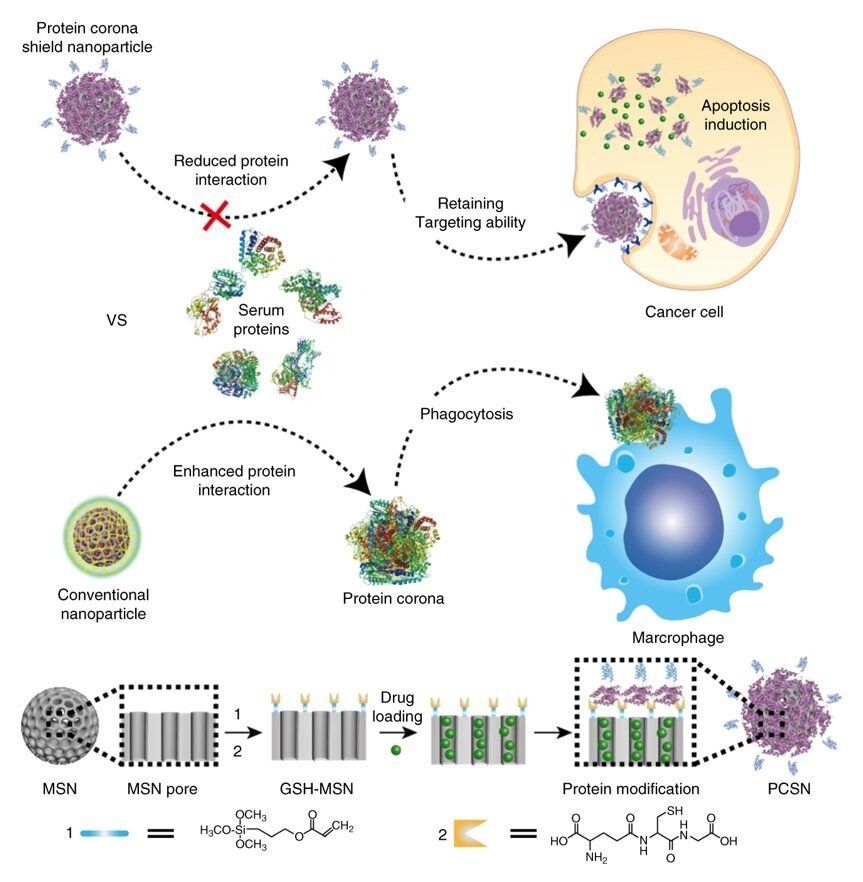
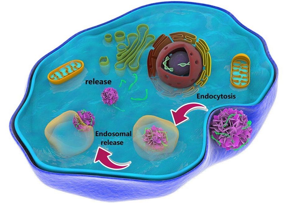
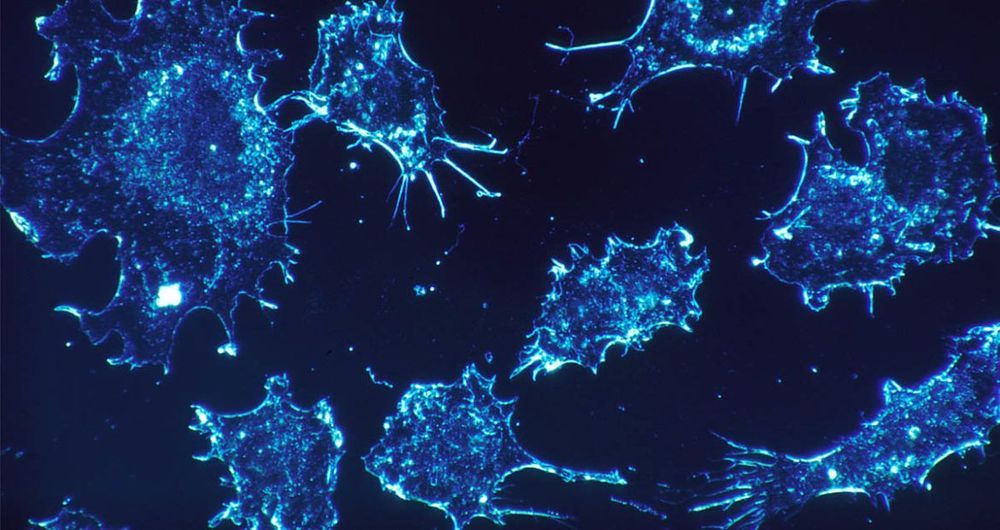
A recent study, affiliated with South Korea’s Ulsan National Institute of Science and Technology (UNIST) has introduced a novel targeted drug delivery system in the fight against cancer.
The past of our ancestors lives on through us: Groundbreaking research illustrates how parental experience is not only epigenetically imprinted onto offspring, but onto an unprecedented number of future generations. Rather than occurring over the elongated time scale of millions of years, genetic change can transpire in real biological time through nanoparticles known as exosomes…
Until recently, it was believed that our genes dictate our destiny. That we are slated for the diseases that will ultimately beset us based upon the pre-wired indecipherable code written in stone in our genetic material. The burgeoning field of epigenetics, however, is overturning these tenets, and ushering in a school of thought where nurture, not nature, is seen to be the predominant influence when it comes to genetic expression and our freedom from or affliction by chronic disease.
Epigenetics: the demise of biological determinism.
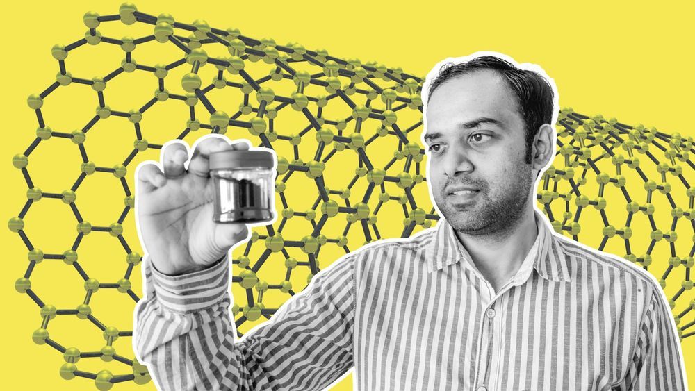
Ever since he was a young boy growing up in Bengaluru, the city home to India’s space research organisation, he dreamt of going to space one day. Now he wants to be the first human on Mars.
One would expect him to train to be an astronaut or dismiss the idea altogether. But that’s not how he plans to get to the red planet.
Meet Gadhadar Reddy, a nano-technologist from Bengaluru, who might just get to live his dream someday in the near future thanks to a new material his company is manufacturing.
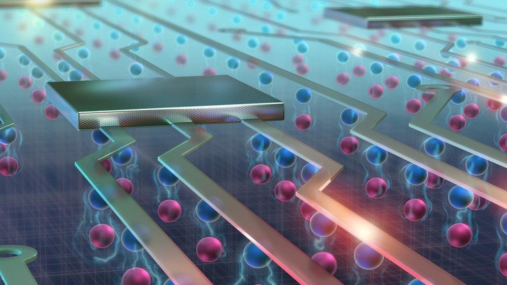
University of New South Wales researchers at the Centre of Excellence for Quantum Computation and Communication Technology (CQC2T) have shown for the first time that they can build atomic precision qubits in a 3D device — another major step towards a universal quantum computer.
The team of researchers, led by 2018 Australian of the Year and Director of CQC2T Professor Michelle Simmons, have demonstrated that they can extend their atomic qubit fabrication technique to multiple layers of a silicon crystal — achieving a critical component of the 3D chip architecture that they introduced to the world in 2015. This new research was published today in Nature Nanotechnology (“Spin read-out in atomic qubits in an all-epitaxial three-dimensional transistor”).
Great Open Access article on Nanotechnology to ring in the new year. #Enjoy
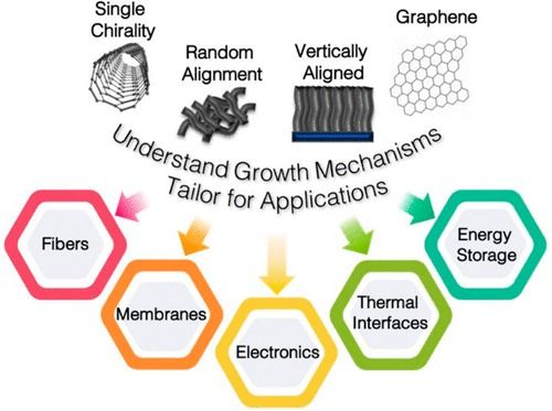
Advances in the synthesis and scalable manufacturing of single-walled carbon nanotubes (SWCNTs) remain critical to realizing many important commercial applications. Here we review recent breakthroughs in the synthesis of SWCNTs and highlight key ongoing research areas and challenges. A few key applications that capitalize on the properties of SWCNTs are also reviewed with respect to the recent synthesis breakthroughs and ways in which synthesis science can enable advances in these applications. While the primary focus of this review is on the science framework of SWCNT growth, we draw connections to mechanisms underlying the synthesis of other 1D and 2D materials such as boron nitride nanotubes and graphene.
2D materials ; boron nitride nanotubes ; carbon nanotubes ; chirality control ; CVD ; graphene ; helicity ; synthesis ;

(Hoboken, N.J. — Nov. 7, 2018) — In their latest feat of engineering, researchers at Stevens Institute of Technology have taken an ordinary white button mushroom from a grocery store and made it bionic, supercharging it with 3D-printed clusters of cyanobacteria that generate electricity and swirls of graphene nanoribbons that can collect the current.
The work, reported in the Nov. 7 issue of Nano Letters, may sound like something straight out of Alice in Wonderland, but the hybrids are part of a broader effort to better improve our understanding of cells biological machinery and how to use those intricate molecular gears and levers to fabricate new technologies and useful systems for defense, healthcare and the environment.
“In this case, our system – this bionic mushroom — produces electricity,” said Manu Mannoor, an assistant professor of mechanical engineering at Stevens. “By integrating cyanobacteria that can produce electricity, with nanoscale materials capable of collecting the current, we were able to better access the unique properties of both, augment them, and create an entirely new functional bionic system.”
High-energy X-ray beams and a clever experimental setup allowed researchers to watch a high-pressure, high-temperature chemical reaction to determine for the first time what controls formation of two different nanoscale crystalline structures in the metal cobalt. The technique allowed continuous study of cobalt nanoparticles as they grew from clusters including tens of atoms to crystals as large as five nanometers.
After developing a method to control exciton flows at room temperature, EPFL scientists have discovered new properties of these quasiparticles that can lead to more energy-efficient electronic devices.
They were the first to control exciton flows at room temperature. And now, the team of scientists from EPFL’s Laboratory of Nanoscale Electronics and Structures (LANES) has taken their technology one step further. They have found a way to control some of the properties of excitons and change the polarization of the light they generate. This can lead to a new generation of electronic devices with transistors that undergo less energy loss and heat dissipation. The scientists’ discovery forms part of a new field of research called valleytronics and has just been published in Nature Photonics.
Excitons are created when an electron absorbs light and moves into a higher energy level, or “energy band” as they are called in solid quantum physics. This excited electron leaves behind an “electron hole” in its previous energy band. And because the electron has a negative charge and the hole a positive charge, the two are bound together by an electrostatic force called a Coulomb force. It’s this electron-electron hole pair that is referred to as an exciton.
A quick and easy test devised by scientists from the University of Queensland could transform cancer diagnosis as we know it.
Cancer is a difficult disease to diagnose because different types are characterised by different signatures. Until now, scientists have been unable to find a unique signature common to all forms of cancer that would set it apart from healthy cells.
That’s what University of Queensland researchers Dr Laura Carrascosa, Dr Abu Sina and Professor Matt Trau have addressed. They have discovered a unique DNA nanostructure that seems to be common to all types of cancer and is visible when cancer cells are placed in water.