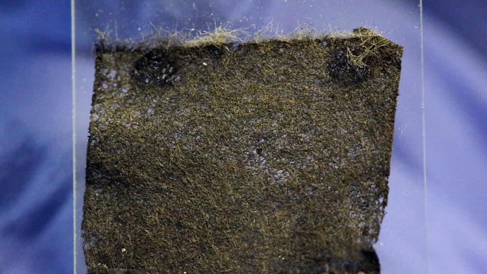
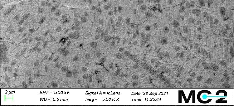
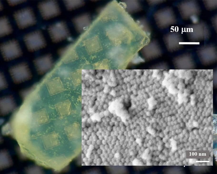
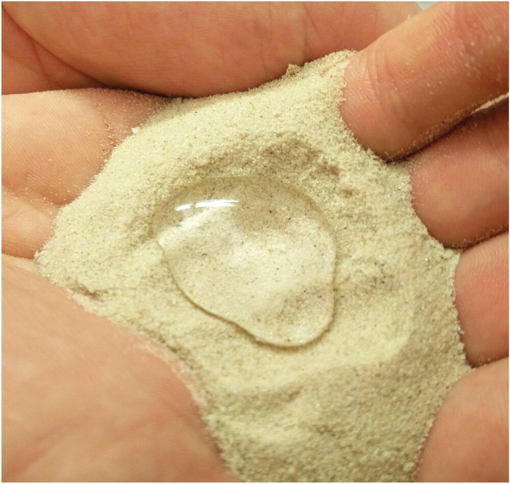
A paint so impractical (and cool) — think matte black squared — you know some idiot will have to try it on a street car, even though it doesn’t like being left outside, or washed. This nanotube coating reflects just 0.036 percent of light.
Category: nanotechnology – Page 222
Researchers at the University of Helsinki in collaboration with researchers from Åbo Akademi University (Finland) and Huazhong University of Science and Technology (China) have developed a new anti-cancer nanomedicine for targeted cancer chemotherapy. This new nano-tool provides a new approach to use cell-based nanomedicines for efficient cancer chemotherapy.
SCIENTISTS have created a “shrink ray” that can reduce objects to one-thousandth of their original size.
The mind-blowing gizmo could one day be used to create nano-robots that are smaller that we can physically produce today.
This brings us a step closer to making 1989 sci-fi comedy Honey, I Shrunk the Kids a reality.
Over the past few years, thermoelectric generators have become the focus of a growing number of studies, due to their ability to convert waste heat into electrical energy. Quantum dots, semiconductor crystals with distinctive conductive properties, could be good candidates for thermoelectric generation, as their discrete resonant levels provide excellent energy filters.
In a recent study, researchers at the University of Cambridge, in collaboration with colleagues in Madrid, Rochester, Duisburg and Sheffield, have experimentally demonstrated the potential of an autonomous nanoscale energy harvester based on resonant tunneling quantum dots. This harvester is based on previous research carried out by part of their team, who had proposed a three-terminal energy harvester based on two resonant-tunneling quantum dots with different energy levels.
The energy harvester device was realized at Cavendish Laboratory in Cambridge by a researcher called Gulzat Jaliel. The original theoretical proposal for the device, however, was introduced by Andrew Jordan in 2013, and the theoretical work behind the harvester was carried out by him in collaboration with renowned semiconductor physicist Markus Büttiker and a team of post-doctoral students in Geneva.
In regenerative medicine, scientists aim to significantly advance techniques that can control stem cell lineage commitment. For example, mechanical stimulation of mesenchymal stem cells (MSCs) at the nanoscale can activate mechanotransduction pathways to stimulate osteogenesis (bone development) in 2-D and 3D culture. Such work can revolutionize bone graft procedures by creating graft material from autologous or allogenic sources of MSCs without chemically inducing the phenomenon. Due to increasing biomedical interest in such mechanical stimulation of cells for clinical use, both researchers and clinicians require a scalable bioreactor system to provide consistently reproducible results. In a new study now published on Scientific Reports, Paul Campsie and a team of multidisciplinary researchers at the departments of biomedical engineering, computing, physics, and molecular, cell and systems biology engineered a new bioreactor system to meet the existing requirements.
The new instrument contained a vibration plate for bioreactions, calibrated and optimized for nanometer vibrations at 1 kHz, a power supply unit to generate a 30 nm vibration amplitude and custom six-well cultureware for cell growth. The cultureware contained magnetic inserts to attach to the bioreactor’s magnetic vibration plate. They assessed osteogenic protein expression to confirm the differentiation of MSCs after initial biological experiments within the system. Campsie et al. conducted atomic force microscopy (AFM) of the 3D gel constructs to verify that strain hardening of the gel did not occur during vibrational stimulation. The results confirmed cell differentiation to be the result of nano-vibrational stimulations provided by the bioreactor alone.
The increasing incidence of skeletal injuries due to age-related conditions such as osteoporosis and osteoarthritis is a metric of the depleting quality of human life. The development of treatments for increased bone density or fracture healing are prime targets for the regenerative potential of mesenchymal stem cells (MSCs). Researchers have demonstrated controlled osteogenesis (development of bones) of MSCs via mechanical stimulation using several methods, including passive and active strategies. Passive methods typically alter the substrate topography to influence the cell adhesion profile, while active methods include exposure to varied forces from external sources.
If used to make non-heritable genetic changes, CRISPR gene-editing technology holds tremendous promise for treating or curing a wide range of devastating disorders, including sickle cell disease, vision loss, and muscular dystrophy. Early efforts to deliver CRISPR-based therapies to affected tissues in a patient’s body typically have involved packing the gene-editing tools into viral vectors, which may cause unwanted immune reactions and other adverse effects.
Now, NIH-supported researchers have developed an alternative CRISPR delivery system: nanocapsules. Not only do these tiny, synthetic capsules appear to pose a lower risk of side effects, they can be precisely customized to deliver their gene-editing payloads to many different types of cells or tissues in the body, which can be extremely tough to do with a virus. Another advantage of these gene-editing nanocapsules is that they can be freeze-dried into a powder that’s easier than viral systems to transport, store, and administer at different doses.
In findings published in Nature Nanotechnology [1], researchers, led by Shaoqin Gong and Krishanu Saha, University of Wisconsin-Madison, developed the nanocapsules with specific design criteria in mind. They would need to be extremely small, about the size of a small virus, for easy entry into cells. Their surface would need to be adaptable for targeting different cell types. They also had to be highly stable in the bloodstream and yet easily degraded to release their contents once inside a cell.
When it comes to finding new treatments for cancer scientitists have been focusing on an anti-cancer agent known as Small interfering ribonucleic acid (siRNA). But getting this agent to cancer cells has been a challenge.
Scientists have developed a platform using nanoparticles to send a cancer-fighting agent to cells.
Scientists from the University of Cambridge have developed a platform that uses nanoparticles known as metal-organic frameworks to deliver a promising anti-cancer agent to cells.
Research led by Dr. David Fairen-Jimenez, from the Cambridge Department of Chemical Engineering and Biotechnology, indicates metal-organic frameworks (MOFs) could present a viable platform for delivering a potent anti-cancer agent, known as siRNA, to cells.
Small interfering ribonucleic acid (siRNA), has the potential to inhibit overexpressed cancer-causing genes, and has become an increasing focus for scientists on the hunt for new cancer treatments.
The replacement of animals as test subjects is one step closer to reality with the successful testing of multi-organ “human-on-a-chip” models to recapitulate the 28-day experiments typically used in animals to evaluate the systemic toxicity of drug and cosmetic compounds. As published and featured as a frontispiece in the prestigious peer-reviewed scientific journal Advanced Functional Materials, the microfluidic device with interlinking modules containing human-derived heart, liver, skeletal muscle and nervous system cells was able to maintain cellular viability and record cellular function in real-time for 28 days.
The University of Central Florida (UCF) in collaboration with the Florida biotech firm Hesperos, Inc., has shown that one of its innovative four-organ in vitro (out of body) model systems is able to realistically replicate in vivo (in body) responses to sustained drug dosing of human cells.
“The technology could allow us, in the very near future, to move chronic drug experiments from animal models to these novel human in vitro models,” said Hesperos Chief Scientist James J. Hickman, who is a Professor at UCF’s NanoScience Technology Center.
A new nanomaterial developed by scientists at the University of Bath could solve a conundrum faced by scientists probing some of the most promising types of future pharmaceuticals.
Scientists who study the nanoscale—with molecules and materials 10,000 smaller than a pinhead—need to be able to test the way that some molecules twist, known as their chirality, because mirror image molecules with the same structure can have very different properties. For instance one kind of molecule smells of lemons when it twists in one direction, and oranges when twisted the other way.
Detecting these twists is especially important in some high-value industries such as pharmaceuticals, perfumes, food additives and pesticides.