Over the past several years, the increased application of unmanned air vehicles (UAVs) in a wide variety of industries has inspired both public and private research laboratories to not only continually improve this technology, but to also support the miniaturization of these devices. The development of both micro- and nano-UAVs is directly related to the ability of researchers to miniaturize the major components of these devices, some of which include micro-processors, sensors, batteries and all necessary wireless communication units that allow UAVs to function properly in any given settings.
Category: nanotechnology – Page 204
With the help of the U.S. Department of Energy’s (DOE) Brookhaven National Laboratory, Juliette Stecenko is exploring cosmology—a branch of astronomy that investigates the origin and evolution of the universe, from the Big Bang to today and into the future. As an intern through DOE’s Science Undergraduate Laboratory Internships (SULI) program, administered at Brookhaven by the Office of Educational Programs (OEP), Stecenko is using modern supercomputers and quantum computing platforms to perform astronomy simulations that may help us better understand where we came from.
Stecenko works under the guidance of Michael McGuigan, a computational scientist in the quantum computing group at Brookhaven’s Computational Science Initiative. The two have been collaborating on simulating Casimir energy—a small force that two electrically neutral surfaces held a tiny distance apart will experience from quantum, atomic, or subatomic fluctuations in the vacuum of space. The vacuum energy of the universe and the Casimir pressure of this energy could be a possible explanation of the origin and evolution of the universe, as well a possible cause of its accelerated expansion.
“Casimir energy is something scientists can measure in the laboratory and is especially important for nanoscience, or in cosmology, in the very early universe when the universe was very small,” McGuigan said.
Carbon nanotubes embedded in leaves detect chemical signals that are produced when a plant is damaged.
MIT engineers have developed a way to closely track how plants respond to stresses such as injury, infection, and light damage, using sensors made of carbon nanotubes. These sensors can be embedded in plant leaves, where they report on hydrogen peroxide signaling waves.
Plants use hydrogen peroxide to communicate within their leaves, sending out a distress signal that stimulates leaf cells to produce compounds that will help them repair damage or fend off predators such as insects. The new sensors can use these hydrogen peroxide signals to distinguish between different types of stress, as well as between different species of plants.
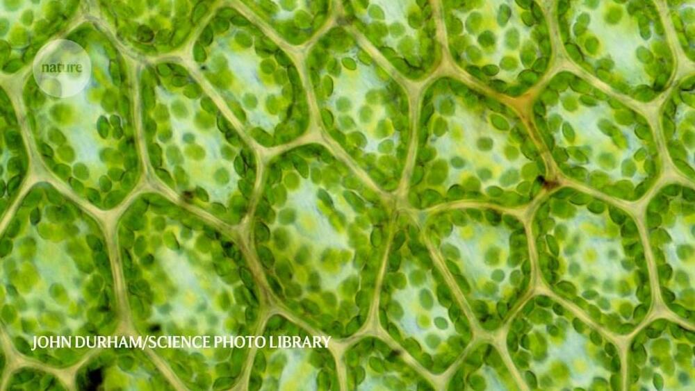
Over billions of years, microorganisms and plants evolved the remarkable process we know as photosynthesis. Photosynthesis converts sun energy into chemical energy, thus providing all life on Earth with food and oxygen. The cellular compartments housing the molecular machines, the chloroplasts, are probably the most important natural engines on earth. Many scientists consider artificially rebuilding and controlling the photosynthetic process the “Apollo project of our time.” It would mean the ability to produce clean energy—clean fuel, clean carbon compounds such as antibiotics, and other products simply from light and carbon dioxide.
But how to build a living, photosynthetic cell from scratch? Key to mimicking the processes of a living cell is to get its components to work together at the right time and place. At the Max Planck Society, this ambitious goal is pursued in an interdisciplinary multi-lab initiative, the MaxSynBio network. Now the Marburg research team led by director Tobias Erb has succeeded successfully created a platform for the automated construction of cell-sized photosynthetically active compartments, “artificial chloroplasts,” that are able to capture and convert the greenhouse gas carbon dioxide with light.
Writing in the journal NanoResearch, a team at the University of Massachusetts Amherst reports this week that they have developed bioelectronic ammonia gas sensors that are among the most sensitive ever made.
The sensor uses electric-charge-conducting protein nanowires derived from the bacterium Geobacter to provide biomaterials for electrical devices. More than 30 years ago, senior author and microbiologist Derek Lovley discovered Geobacter in river mud. The microbes grow hair-like protein filaments that work as nanoscale “wires” to transfer charges for their nourishment and to communicate with other bacteria.
First author and biomedical engineering doctoral student Alexander Smith, with his advisor Jun Yao and Lovley, say they designed this first sensor to measure ammonia because that gas is important to agriculture, the environment and biomedicine. For example, in humans, ammonia on the breath may signal disease, while in poultry farming, the gas must be closely monitored and controlled for bird health and comfort and to avoid feed imbalances and production losses.
Ben-Gurion University of the Negev (BGU) researchers are developing safe anti-viral nanoparticle coatings that demonstrate significant potential in preventing active surface infection with SARS-CoV-2.
Some days I feel like I’m living under a rock. How did I miss the development of a superfast nanobot that is controlled by exterior electric fields? This is an extraordinary development.
Piecework at the nano assembly line.
Quantum technology is currently one of the most active fields of research worldwide. It takes advantage of the special properties of quantum mechanical states of atoms, light, or nanostructures to develop, for example, novel sensors for medicine and navigation, networks for information processing and powerful simulators for materials sciences. Generating these quantum states normally requires a strong interaction between the systems involved, such as between several atoms or nanostructures.
Until now, however, sufficiently strong interactions were limited to short distances. Typically, two systems had to be placed close to each other on the same chip at low temperatures or in the same vacuum chamber, where they interact via electrostatic or magnetostatic forces. Coupling them across larger distances, however, is required for many applications such as quantum networks or certain types of sensors.
A team of physicists, led by Professor Philipp Treutlein from the Department of Physics at the University of Basel and the Swiss Nanoscience Institute (SNI), has now succeeded for the first time in creating strong coupling between two systems over a greater distance across a room temperature environment. In their experiment, the researchers used laser light to couple the vibrations of a 100 nanometer thin membrane to the motion of the spin of atoms over a distance of one meter. As a result, each vibration of the membrane sets the spin of the atoms in motion and vice versa.
Nanotechnology is the application of science at a truly nano scale. To put that in perspective, if a nanometre were the size of a cup of tea, a meter would cover the diameter of the whole Earth.
Being able to control the world at such an intricate level has the potential to revolutionise medicine – enabling us to target cancer cells, deliver drugs and fight antibiotic resistance – but how do we create technology to that size?
Sonia talks to our editorial assistant Amy Barret about how her work in nanotechnology began, building proteins unknown to nature, and why going nano is nothing like in the movies.
Researchers at ARCNL and Vrije Universiteit Amsterdam have developed a compact setup for fast, super-resolution microscopy through an ultrathin fiber. Using smart signal processing, they beat the theoretical limits of resolution and speed. Because the method does not require any special fluorescent labelling, it is promising for both medical applications and characterization of 3D structures in nanolithography. On May 7th, the results were published in Light: Science & Applications, a scientific journal in the Nature family.
“Imaging at the nanoscale is limited by the wavelength of the light that is used. There are ways to overcome this diffraction limit, but they typically require large microscopes and difficult processing procedures,” says Lyuba Amitonova. “These systems are unsuitable for imaging in deep layers of biological tissue or in other hard-to-reach places.”
Amitonova recently started a research group on Nanoscale Imaging and Metrology at ARCNL. She is also connected part-time to VU Amsterdam where she works on ultrathin fibers for endomicroscopy in the group of Johannes de Boer. Amitonova and de Boer have developed a way to overcome the diffraction limit in small systems to enable deep-tissue imaging with super-resolution.