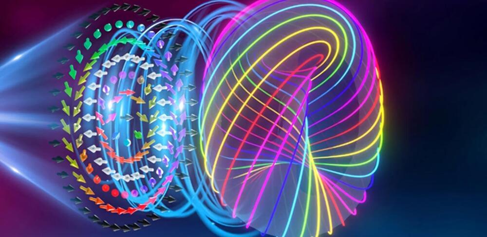
Toggle light / dark theme

Here’s a new chapter in the story of the miniaturisation of machines: researchers in a laboratory in Singapore have shown that a single atom can function as either an engine or a fridge. Such a device could be engineered into future computers and fuel cells to control energy flows.

“Think about how your computer or laptop has a lot of things inside it that heat up. Today you cool that with a fan that blows air. In nanomachines or quantum computers, small devices that do cooling could be something useful,” says Dario Poletti from the Singapore University of Technology and Design (SUTD).

This work gives new insight into the mechanics of such devices. The work is a collaboration involving researchers at the Centre for Quantum Technologies (CQT) and Department of Physics at the National University of Singapore (NUS), SUTD and at the University of Augsburg in Germany. The results were published in the peer-reviewed journal npj Quantum Information on 1 May.

HOUSTON, May 1, 2020 — Rice University researchers plan to reconfigure their wastewater-treatment technology to capture and deactivate the virus that causes COVID-19. Their chemical-free nanotechnology, introduced earlier this year as a way to kill bacterial “superbugs” and degrade their antibiotic resistance genes in wastewater, will use graphitic carbon nitride to selectively adsorb viruses and then disable them by activating nearby catalysts with light. The team believes that this photocatalytic approach to disinfection — what it calls the “trap-and-zap” treatment approach — could be used to recognize coronaviruses that cause not only COVID-19 but also MERS and SARS.

North­eastern Uni­ver­sity and Wen­zhou Med­ical Uni­ver­sity in China have part­nered to com­bine their exper­tise in nan­otech­nology and med­i­cine, respec­tively, to col­lab­o­rate on research focusing on reducing eye infections caused by contact lenses and intraocular devices.

Through this part­ner­ship, the researchers will explore the emerging field of picotech­nology, which involves the manip­u­la­tion of matter on an even smaller scale than is used in nanotechnology.


Nanotechnology researchers at Northeastern are joining forces with Wenzhou Medical University in China to collaborate in the new field of picotechnology—and combat eye infections.


Not long ago nanotechnology was a fringe topic; now it’s a flourishing engineering field, and fairly mainstream. For example, while writing this article, I happened to receive an email advertisement for the “Second World Conference on Nanomedicine and Drug Delivery,” in Kerala, India. It wasn’t so long ago that nanomedicine seemed merely a flicker in the eyes of Robert Freitas and a few other visionaries!

But nano is not as small as the world goes. A nanometer is 10−9 meters – the scale of atoms and molecules. A water molecule is a bit less than one nanometer long, and a germ is around a thousand nanometers across. On the other hand, a proton has a diameter of a couple femtometers – where a femtometer, at 10−15 meters, makes a nanometer seem positively gargantuan. Now that the viability of nanotech is widely accepted (in spite of some ongoing heated debates about the details), it’s time to ask: what about femtotech? Picotech or other technologies at the scales between nano and femto seem relatively uninteresting, because we don’t know any basic constituents of matter that exist at those scales. But femtotech, based on engineering structures from subatomic particles, makes perfect conceptual sense, though it’s certainly difficult given current technology.

The nanotech field was arguably launched by Richard Feynman’s 1959 talk “There’s Plenty of Room at the Bottom.” As Feynman wrote there.

Alzheimer’s disease is the sixth leading cause of death in the United States, affecting one in 10 people over the age of 65. Scientists are engineering nanodevices to disrupt processes in the brain that lead to the disease.

People who are affected by Alzheimer’s disease have a specific type of plaque, made of self-assembled molecules called β-amyloid (Aβ) , that build up in the brain over time. This buildup is thought to contribute to loss of neural connectivity and . Researchers are studying ways to prevent the peptides from forming these dangerous plaques in order to halt development of Alzheimer’s disease in the brain.

In a multidisciplinary study, scientists at the U.S. Department of Energy’s (DOE) Argonne National Laboratory, along with collaborators from the Korean Institute of Science and Technology (KIST) and the Korea Advanced Institute of Science and Technology (KAIST), have developed an approach to prevent plaque formation by engineering a nano-sized device that captures the dangerous peptides before they can self-assemble.

Researchers have demonstrated the world’s first metasurface laser that produces “super-chiral light”: light with ultra-high angular momentum. The light from this laser can be used as a type of “optical spanner” to or for encoding information in optical communications.

“Because can carry angular , it means that this can be transferred to matter. The more angular momentum light carries, the more it can transfer. So you can think of light as an ‘optical spanner’,” Professor Andrew Forbes from the School of Physics at the University of the Witwatersrand (Wits) in Johannesburg, South Africa, who led the research. “Instead of using a physical spanner to twist things (like screwing nuts), you can now shine light on the nut and it will tighten itself.”

The new produces a new high purity “twisted light” not observed from lasers before, including the highest angular momentum reported from a laser. Simultaneously the researchers developed a nano-structured that has the largest phase gradient ever produced and allows for high power operation in a compact design. The implication is a world-first laser for producing exotic states of twisted structured light, on demand.

Existing electronic skin (e-skin) sensing platforms are equipped to monitor physical parameters using power from batteries or near-field communication. For e-skins to be applied in the next generation of robotics and medical devices, they must operate wirelessly and be self-powered. However, despite recent efforts to harvest energy from the human body, self-powered e-skin with the ability to perform biosensing with Bluetooth communication are limited because of the lack of a continuous energy source and limited power efficiency. Here, we report a flexible and fully perspiration-powered integrated electronic skin (PPES) for multiplexed metabolic sensing in situ. The battery-free e-skin contains multimodal sensors and highly efficient lactate biofuel cells that use a unique integration of zero- to three-dimensional nanomaterials to achieve high power intensity and long-term stability. The PPES delivered a record-breaking power density of 3.5 milliwatt·centimeter−2 for biofuel cells in untreated human body fluids (human sweat) and displayed a very stable performance during a 60-hour continuous operation. It selectively monitored key metabolic analytes (e.g., urea, NH4+, glucose, and pH) and the skin temperature during prolonged physical activities and wirelessly transmitted the data to the user interface using Bluetooth. The PPES was also able to monitor muscle contraction and work as a human-machine interface for human-prosthesis walking.

Recent advances in robotics have enabled soft electronic devices at different scales with excellent biocompatibility and mechanical properties; these advances have rendered novel robotic functionalities suitable for various medical applications, such as diagnosis and drug delivery, soft surgery tools, human-machine interaction (HMI), wearable computing, health monitoring, assistive robotics, and prosthesis (1–6). Electronic skin (e-skin) can have similar characteristics to human skin, such as mechanical durability and stretchability and the ability to measure various sensations such as temperature and pressure (7–11). Moreover, e-skin can be augmented with capabilities beyond those of the normal human skin by incorporating advanced bioelectronics materials and devices.

Researchers have developed a number of potassium ion (K+) probes to detect fluctuating K+ concentrations during a variety of biological processes. However, such probes are not sensitive enough to detect physiological fluctuations in living animals and it is not easy to monitor deep tissues with short-wavelength excitations that are in use so far. In a new report, Jianan Liu and a team of researchers in neuroscience, chemistry, and molecular engineering in China, describe a highly sensitive and selective nanosensor for near infrared (NIR) K+ ion imaging in living cells and animals. The team constructed the nanosensor by encapsulating upconversion nanoparticles (UCNPs) and a commercial potassium ion indicator in the hollow cavity of mesoporous silica nanoparticles and coated them with a K+ selective filter membrane. The membrane adsorbed K+ from the medium and filtered away any interfering cations. In its mechanism of action, UCNPs converted NIR to ultraviolet (UV) light to excite the potassium ion indicator and detect fluctuating potassium ion concentrations in cultured cells and in animal models of disease including mice and zebrafish larvae. The results are now published on Science Advances.

The most abundant intracellular cation potassium (K+) is extremely crucial in a variety of biological processes including neural transmission, heartbeat, muscle contraction and kidney function. Variations in the intracellular or extracellular K+ concentration (referred herein as [K+]) suggest abnormal physiological functions including heart dysfunction, cancer, and diabetes. As a result, researchers are keen to develop effective strategies to monitor the dynamics of [K+] fluctuations, specifically with direct optical imaging.

Most existing probes are not sensitive to K+ detection under physiological conditions and cannot differentiate fluctuations between [K+] and the accompanying sodium ion ([Na+]) during transmembrane transport in the Na+/K+ pumps. While fluorescence lifetime imaging can distinguish K+ and Na+ in water solution, the method requires specialized instruments. Most K+ sensors are also activated with short wavelength light including ultraviolet (UV) or visible light—leading to significant scattering and limited penetration depth when examining living tissues. In contrast, the proposed near-infrared (NIR) imaging technique will offer unique advantages during deep tissue imaging as a plausible alternative.

Researchers investigating cribellate spiders have discovered a unique comb structure that could help inform future equipment used to manipulate nanofibers. Nanofibers have been hard to handle in a lab setting as they can stick to the equipment attempting to manipulate them, but a new study published in the journal ACS Applied Nanomaterials reveals how spiders can help us to create non-stick tools for such scenarios.

Cribellate spiders are so named because of their unique web-spinning anatomy. Most spiders have a long single spinneret that they use to produce a single thread, whereas cribellate spiders have a silk-spinning organ. This organ acts like a plate with lots of small, ever so slightly raised protrusions, each of which produces a very fine silk just a few nanometers thick. The spiders then comb these thin fibers out using a calamistrum structure on their legs, producing silk with a woolly texture. This woolly-textured silk entraps the spider’s prey, but somehow, they are able to handle it without getting caught up in their own webs.

Nanofibers are a hot area of research right now but one of the difficulties in their handling is that they commonly stick to the equipment trying to manipulate them. Lead author Anna-Christin Joel, from RWTH Aachen University, and her colleagues wondered if the solution to this frustrating problem could be found within the silk-immune spiders’ anatomy.

Engineering researchers developed a next-generation miniature lab device that uses magnetic nano-beads to isolate minute bacterial particles that cause diseases. Using this new technology improves how clinicians isolate drug-resistant strains of bacterial infections and difficult-to-detect micro-particles such as those making up Ebola and coronaviruses.

Ke Du and Blanca Lapizco-Encinas, both faculty-researchers in Rochester Institute of Technology’s Kate Gleason College of Engineering, worked with an international team to collaborate on the design of the new system — a microfluidic device, essentially a lab-on-a-chip.

Drug-resistant bacterial infections are causing hundreds of thousands of deaths around the world every year, and this number is continuously increasing. Based on a report from the United Nations, the deaths caused by antibiotics resistance could reach to 10 million annually by 2050, Du explained.