Toggle light / dark theme

Researchers have significantly accelerated ion movement using nanotechnology, potentially improving technologies from battery charging to biosensing.

This breakthrough at Washington State University and Lawrence Berkeley National Laboratory involves creating a nanochannel lined with molecules that attract ions, allowing them to move over ten times faster than before. This development could revolutionize energy storage and help detect environmental pollutants or neurological activities.

Breaking Speed Records With Nanoscience

A new method enables researchers to analyze magnetic nanostructures with a high resolution. It was developed by researchers at Martin Luther University Halle-Wittenberg (MLU) and the Max Planck Institute of Microstructure Physics in Halle.

The new method achieves a resolution of around 70 nanometers, whereas normal light microscopes have a resolution of just 500 nanometers. This result is important for the development of new, energy-efficient storage technologies based on spin electronics. The team reports on its research in the current issue of the journal ACS Nano.

Normal optical microscopes are limited by the wavelength of light and details below around 500 nanometers cannot be resolved. The new method overcomes this limit by utilizing the anomalous Nernst effect (ANE) and a metallic nano-scale tip. ANE generates an electrical voltage in a magnetic metal that is perpendicular to the magnetization and a .

Researchers from Seoul National University College of Engineering announced they have developed an optical design technology that dramatically reduces the volume of cameras with a folded lens system utilizing “metasurfaces,” a next-generation nano-optical device.

By arranging metasurfaces on the so that light can be reflected and moved around in the glass substrate in a folded manner, the researchers have realized a with a thickness of 0.7mm, which is much thinner than existing refractive lens systems. The research was published on Oct. 30 in the journal Science Advances.

Traditional cameras are designed to stack multiple glass lenses to refract light when capturing images. While this structure provided excellent high-quality images, the thickness of each lens and the wide spacing between lenses increased the overall bulk of the camera, making it difficult to apply to devices that require ultra-compact cameras, such as virtual and augmented reality (VR-AR) devices, smartphones, endoscopes, drones, and more.

Tissues take shape during development through a series of morphogenetic movements guided by local cell-scale forces. While current in vitro approaches subjecting tissues to homogenous stresses, it is currently no possible to recapitulate highly local spatially varying forces. Here we develop a method for local actuation of organoids using embedded magnetic nanoparticles. Sequential aggregation of magnetically labelled human pluripotent stem cells followed by actuation by a magnetic field produces localized magnetic clusters within the organoid. These clusters impose local mechanical forces on the surrounding tissue in response to applied global magnetic fields. We show that precise, spatially defined actuation provides short-term mechanical tissue perturbations as well as long-term cytoskeleton remodeling. We demonstrate that local magnetically-driven actuation guides asymmetric growth and proliferation, leading to enhanced patterning in human neural organoids. We show that this approach is applicable to other model systems by observing polarized patterning in paraxial mesoderm organoids upon local magnetic actuation. This versatile approach allows for local, controllable mechanical actuation in multicellular constructs, and is widely applicable to interrogate the role of local mechanotransduction in developmental and disease model systems.

The authors have declared no competing interest.

From repairing deadly brain bleeds to tackling tumors with precise chemotherapy, micro/nano-robots (MNRs) are a promising, up-and-coming tool that have the power to substantially advance health care. However, this tool still has difficulty navigating within the human body—a limitation that has prevented it from entering clinical trials.

Mathematical models are crucial to the optimal design and navigation of MNRs, but the are inadequate. Now, new, promising research from the University of Saskatchewan (USask) may allow MNRs to overcome the limitations that previously prevented their widespread use.

USask College of Engineering professor Dr. Chris Zhang (Ph. D.) and two Ph.D. students (Lujia Ding, N.N Hu) along with two USask alumni (Dr. Bing Zhang (Ph. D.), Dr. R. Y. Yin (Ph. D.)) are the first team to develop a highly accurate mathematical model that optimizes the design of MNRs which improves their navigation, allowing them to travel efficiently through the bloodstream. Their work was recently published in Nature Communications.

Surfaces play a key role in numerous chemical reactions, including catalysis and corrosion. Understanding the atomic structure of the surface of a functional material is essential for both engineers and chemists. Researchers at Nagoya University in Japan used atomic-resolution secondary electron (SE) imaging to capture the atomic structure of the very top layer of materials to better understand the differences from its lower layers. The researchers published their findings in the journal Microscopy.

Some materials exhibit “surface reconstruction,” where the surface atoms are organized differently from the interior atoms. To observe this, especially at the atomic level, surface-sensitive techniques are needed.

Traditionally, scanning (SEM) has been an effective tool to examine nanoscale structures. SEM works by scanning a sample with a focused electron beam and capturing the SEs emitted from the surface. SEs are typically emitted from a below the surface, making it difficult to observe phenomena like surface reconstruction, especially if only a single atomic layer is involved.

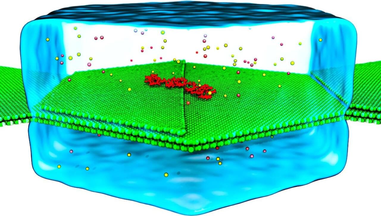
Researchers in the Nanoscience Center at the University of Jyväskylä, Finland, have used machine learning and supercomputer simulations to investigate how tiny gold nanoparticles bind to blood proteins. The studies discovered that favorable nanoparticle-protein interactions can be predicted from machine learning models that are trained from atom-scale molecular dynamics simulations. The new methodology opens ways to simulate the efficacy of gold nanoparticles as targeted drug delivery systems in precision nanomedicine.

Hybrid nanostructures between biomolecules and inorganic nanomaterials constitute a largely unexplored field of research, with the potential for novel applications in bioimaging, biosensing, and nanomedicine. Developing such applications relies critically on understanding the dynamical properties of the nano–bio interface.

Modeling the properties of the nano-bio interface is demanding since the important processes such as electronic charge transfer, or restructuring of the biomolecule surface can take place in a wide range of length and time scales, and the atomistic simulations need to be run in the appropriate aqueous environment.

Join us at.

#AI #ExponentialTechnologies


The book “Intelciety. Intelligent Society. Are We Ready for the Challenge?” explores the profound changes that artificial intelligence (AI) and other emerging technologies are causing in modern society. Vicente Ferreira da Silva addresses how these technologies are transforming various fields, from medicine and biotechnology to robotics and nanotechnology, and questions whether we are truly prepared to deal with these advances.