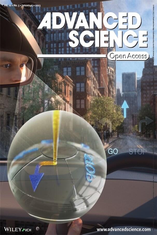
Toggle light / dark theme

NIH launches clinical trial of universal influenza vaccine candidate

A first-in-human, Phase 1 trial assessing the safety and immunogenicity of an investigational nanoparticle influenza vaccine designed to provide long-lasting protection against multiple flu virus strains has begun at the National Institutes of Health Clinical Center in Bethesda, Maryland. Healthy participants 18 to 50 years old will receive either a licensed seasonal influenza vaccine or the experimental vaccine, FluMos-v1. Scientists from NIH’s National Institute of Allergy and Infectious Diseases (NIAID) developed FluMos-v1 to stimulate antibodies against multiple influenza virus strains by displaying part of the influenza virus hemagglutinin (HA) protein on self-assembling nanoparticle scaffolds. Alicia T. Widge, M.D., of NIAID’s Vaccine Research Center (VRC), is the principal investigator of the NIAID-sponsored single-site trial.

“The health and economic burdens of influenza are substantial, and the world badly needs improved flu vaccines,” said NIAID Director Anthony S. Fauci, M.D. “I am encouraged by the great promise of the VRC nanoparticle vaccine candidate, which so far has performed very well in pre-clinical testing.”

Standard influenza vaccines must be reformulated and administered annually to match changes in the HA protein in the viral strains predicted to dominate in the upcoming influenza season. If the vaccine is not well matched to dominant circulating virus strains, the antibodies elicited may provide sub-optimal protection. So-called universal influenza vaccines are being developed and tested by many research groups and could one day eliminate the need for annual vaccination by generating long-lasting antibodies to protect against many existing or emergent influenza virus strains, including those not represented in the vaccine.

U.S. Transhumanist Party Virtual Enlightenment Salon with Ryan O’Shea — May 30, 2021

Today, Sunday, May 30, 2021, at 1 p.m. Pacific Time, join us for a U.S. Transhumanist Party Virtual Enlightenment Salon with Ryan O’Shea, as we discuss the state of the transhumanist movement, life-extension advocacy, biohacking, Ryan’s Future Grind podcast, and more!

Watch on YouTube here:. You will be able to post questions and comments in the live YouTube chat.


On Sunday, May 30, 2021, at 1 p.m. U.S. Pacific Time, the U.S. Transhumanist Party invites Ryan O’Shea for a Virtual Enlightenment Salon to discuss a wide array of subjects related to transhumanism, including the state of the contemporary transhumanist movement, Ryan O’Shea’s Future Grind podcast, biohacking, the Human Augmentation Institute and the Human Augmentation Code of Ethics, Ryan O’Shea’s media work with the Lifespan Extension Advocacy Foundation with the goal of popularizing life-extension science, how to respond to common criticisms of transhumanism, thoughts on consciousness and free will, and strategies for advancing the transhumanist movement in the future.

Ryan O’Shea is an entrepreneur and futurist speaker from Pittsburgh, Pennsylvania. He is the host of Future Grind — https://futuregrind.org/ — a multimedia production company that seeks to increase technoliteracy and democratize access to information about emerging technologies, enabling more voices to be a part of the societal conversation surrounding technology. Ryan is also a founder of the Human Augmentation Institute, an organization focused on upholding bodily autonomy and ensuring that any efforts in human augmentation are done ethically, safely, and responsibly. He also serves as the spokesperson for Grindhouse Wetware, a group specializing in technology to augment human capabilities. In 2017, Ryan co-founded a National Institutes of Health and National Science Foundation-supported artificial intelligence startup that is working to use machine learning and automated just-in-time intervention for behavior change. Ryan has represented NASA and CalTech’s Jet Propulsion Laboratory as a Solar System Ambassador and serves both as a World Economic Forum Global Shaper and an ambassador for Pittsburgh AI. He is a graduate of the University of Pittsburgh and currently serves on the boards of multiple non-profit organizations.

Become a member of the U.S. Transhumanist Party for free, no matter where you reside: https://transhumanist-party.org/membership.

New tool activates deep brain neurons

Using a mouse model, Chen and the team delivered a viral construct containing TRPV1 ion channels to genetically-selected neurons. Then, they delivered small burst of heat via low-intensity focused ultrasound to the select neurons in the brain via a wearable device. The heat, only a few degrees warmer than body temperature, activated the TRPV1 ion channel, which acted as a switch to turn the neurons on or off.


Neurological disorders such as Parkinson’s disease and epilepsy have had some treatment success with deep brain stimulation, but those require surgical device implantation. A multidisciplinary team at Washington University in St. Louis has developed a new brain stimulation technique using focused ultrasound that is able to turn specific types of neurons in the brain on and off and precisely control motor activity without surgical device implantation.

The team, led by Hong Chen, assistant professor of biomedical engineering in the McKelvey School of Engineering and of radiation oncology at the School of Medicine, is the first to provide direct evidence showing noninvasive, cell-type-specific activation of neurons in the brain of mammal by combining ultrasound-induced heating effect and genetics, which they have named sonothermogenetics. It is also the first work to show that the ultrasound-genetics combination can robustly control behavior by stimulating a specific target deep in the brain.

Results of the three years of research, which was funded in part by the National Institutes of Health’s BRAIN Initiative, were published online in Brain Stimulation May 11, 2021.

The next pandemic: Rift Valley fever?

Rift Valley fever used to mostly affect livestock in Africa. But the virus that causes it is also spread by mosquitoes whose habitats are expanding because of climate change. If it were to make its way to the rest of the world, it would decimate livestock causing agricultural collapse as well as affecting human health.

In 2015 the Zika virus triggered a global health crisis that left thousands of parents devastated. The virus can cause serious problems in pregnancy, leading to babies with birth defects called microcephaly and other neurological problems. But Zika is not the only virus that can be devastating to pregnant women and their babies; there is another with pandemic potential that could be even more deadly – Rift Valley fever.

The placenta that encases the baby acts as a fortress against many pathogens, but a few can evade its defences. Rift Valley fever is one of them – a 2019 study shows that the virus has the ability to infect a specialised layer of placental cells that carry nutrients to the baby, something that even Zika may not be capable of. In cattle and other livestock, in which the virus spreads, infection can cause more than 90% of pregnant cows to miscarry or deliver stillborn calves. Although the virus kills fewer than 1% of people it infects, it is the risk to babies, and the lasting neurological effects in adults, that is of great concern.

Dr. Jonathan Chernoff, MD, PhD — Senior VP, Deputy Director, and CSO — Fox Chase Cancer Center

Innovating At The Frontiers Of Cancer Biology — Dr. Jonathan Chernoff MD, PhD, Senior Vice President, Deputy Director, and Chief Scientific Officer, Fox Chase Cancer Center.


Dr. Jonathan Chernoff, MD, PhD, is Senior Vice President, Deputy Director, and Chief Scientific Officer, at Fox Chase Cancer Center (https://www.foxchase.org/) where he coordinates and charts the future course of research for the organization.

The Hospital of Fox Chase Cancer Center and its affiliates (collectively “Fox Chase Cancer Center”), a member of the Temple University Health System, is one of the leading cancer research and treatment centers in the United States. Founded in 1904 in Philadelphia as one of the nation’s first cancer hospitals, Fox Chase was also among the first institutions to be designated a National Cancer Institute Comprehensive Cancer Center in 1974.

Dr. Chernoff joined the staff in 1991 as an associate member and was promoted to member with tenure in 1996. In 2002 he was promoted to be a senior member in Fox Chase Cancer Center’s Basic Science division, the equivalent of a full professor in a university.

A molecular oncologist as well as a board-certified medical oncologist, Dr. Chernoff has a special interest in factors that control cell growth and movement, including oncogenes and anticancer or tumor-suppressor genes, and has made fundamental contributions in this research.

New potential drug target may protect brain against low oxygen damage

Some of the most devastating health effects of a stroke or heart attack are caused by oxygen deprivation in the brain. Now, researchers at Massachusetts General Hospital (MGH) have identified an enzyme that may naturally protect the brain from oxygen deprivation damage, which could be a potential drug target to prevent issues arising from strokes or heart attacks.

Like many scientific breakthroughs, the new discovery came about while investigating something else entirely. The team was looking into a study from 2005 that found that a state of “suspended animation” could be induced in mice by having them inhale hydrogen sulfide. In the new study, the researchers set out to investigate the longer-term effects of that exposure.

The team exposed groups of mice to hydrogen sulfide for four hours a day, for five consecutive days. The suspended animation-like state followed, with the animals’ movement slowing and body temperatures dropping.

The efficacy and safety of Favipiravir in treatment of COVID-19: a systematic review and meta-analysis of clinical trials

The novel coronavirus outbreak began in late December 2019 and rapidly spread worldwide, critically impacting public health systems. A number of already approved and marketed drugs are being tested for repurposing, including Favipiravir. We aim to investigate the efficacy and safety of Favipiravir in treatment of COVID-19 patients through a systematic review and meta-analysis. This systematic review and meta-analysis were reported in accordance with the PRISMA statement. We registered the protocol in the PROSPERO (CRD42020180032). All clinical trials which addressed the safety and efficacy of Favipiravir in comparison to other control groups for treatment of patients with confirmed infection with SARS-CoV2 were included. We searched electronic databases including LitCovid/PubMed, Scopus, Web of Sciences, Cochrane, and Scientific Information Database up to 31 December 2020.

3D printing stem cells to transform neuroscience

3D printing, also called additive manufacturing, has become widespread in recent years. By building successive layers of raw material such as metals, plastics, and ceramics, it has the key advantage of being able to produce very complex shapes or geometries that would be nearly impossible to construct through more traditional methods such as carving, grinding, or molding.

The technology offers huge potential in the health care sector. For example, doctors can use it to make products to match a patient’s anatomy: a radiologist could create an exact replica of a patient’s spine to help plan surgery; a dentist could scan a patient’s broken tooth to make a perfectly fitting crown reproduction. But what if we took a step further and apply 3D printing techniques to neuroscience?

Stems cells are essentially the body’s raw materials; they are pluripotent elements from which all other cells with specialized functions are generated. The development of methods to isolate and generate human stem cells, has excited many with the promise of improved human cell function understanding, ultimately utilizing them for regeneration in disease and trauma. However, the traditional two-dimensional growth of derived neurones–using flat petri dishes–presents itself as a major confounding factor as it does not adequately mimic in vivo three-dimensional interactions, nor the myriad developmental cues present in real living organisms.

To address this limitation in current neuronal culturing approaches, the FET funded MESO-BRAIN project, led by Aston University, proposed a highly ambitious interdisciplinary enterprise to construct truly 3D networks that not only displayed in vivo activity patterns of neural cultures but also allowed for precise interaction with these cultures. This allows the activity of individual elements to be readily monitored and controlled through electrical stimulation.

The ability to develop human-induced pluripotent stem cell derived neural networks upon a defined and reproducible 3D scaffold that can emulate brain activity, allows for a comprehensive and detailed investigation of neural network development.

The MESO-BRAIN project facilitates a better understanding of human disease progression, neuronal growth and enables the development of large-scale human cell-based assays to test the modulatory effects of pharmacological and toxicological compounds on neural network activity. This can ultimately help to better understand and treat neurological conditions such as Parkinson’s disease, dementia, and trauma. In addition, the use of more physiologically relevant human models will increase drug screening efficiency and reduce the need for animal testing.

Highly Pathogenic Bird Flu Outbreak Already Reported in 46 Countries, Scientists Warn

While the world was distracted with the rampant spread of a novel coronavirus, 2020 also witnessed an explosion in another deadly pathogen that could pose a threat to global public health.

H5N8, a subtype of highly pathogenic avian influenza virus (HPAIV), was identified decades ago, but during 2020 a series of emerging and ongoing H5N8 outbreaks in avian populations across dozens of countries have led to the death or slaughter of millions of birds worldwide.

“The affected geographic regions have been expanding continuously, and at least 46 countries have reported highly pathogenic H5N8 AIV outbreaks,” virus researchers Weifeng Shi and George F. Gao write in a new perspective article in Science, warning of the dangers of H5N8 if we don’t closely monitor and contain this worrisome trend.

AI-Powered Smart Toilet May Soon Analyze Poop for Health Problems

Artificial intelligence tool can be used for long-term tracking and management of chronic gastrointestinal ailments.

An artificial intelligence tool under development at Duke University can be added to the standard toilet to help analyze patients’ stool and give gastroenterologists the information they need to provide appropriate treatment, according to research that was selected for presentation at Digestive Disease Week® (DDW) 2021. The new technology could assist in managing chronic gastrointestinal issues such as inflammatory bowel disease (IBD) and irritable bowel syndrome (IBS).

“Typically, gastroenterologists have to rely on patient self-reported information about their stool to help determine the cause of their gastrointestinal health issues, which can be very unreliable,” said Deborah Fisher, MD, one of the lead authors on the study and associate professor of medicine at Duke University Durham, North Carolina. “Patients often can’t remember what their stool looks like or how often they have a bowel movement, which is part of the standard monitoring process. The Smart Toilet technology will allow us to gather the long-term information needed to make a more accurate and timely diagnosis of chronic gastrointestinal problems.”