A new study reveals how viruses that infect bacteria, called bacteriophages or “phages,” use a tiny piece of genetic material to hijack bacterial cells and make more copies of themselves.
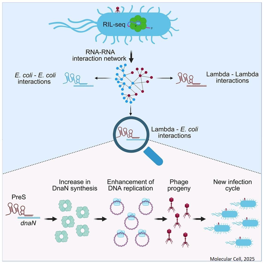
The research shows that a very small RNA molecule, called PreS, acts like a hidden “switch” inside the bacterial cell. By flipping this switch, the virus can change how the bacterial cell works and push the infection forward.
Until now, most phage research has focused on viral proteins. This study shows that phages also use RNA molecules to quickly reprogram the host cell after the bacterial genes have already been read and bacterial messages (mRNAs) were made, adding an extra layer of control during infection.
PreS attaches to these important bacterial messages and tweaks them in a way that helps the virus copy its DNA and move more efficiently toward the stage where new viruses are produced and burst out of the cell, killing the bacterium.
Using advanced methods to map RNA–RNA interactions (termed RIL-seq), the researchers found that one of PreS’s key targets is a bacterial message that makes DnaN, a protein that plays a central role in copying DNA. By helping the cell make more DnaN, PreS gives the virus a strong head start in the infection process.
Interestingly, PreS works by changing the shape of the bacterial dnaN message.
Normally, part of this message is tightly folded, which makes it hard for the cell’s protein-making machines (ribosomes) to access. PreS binds to this folded region, opens it up, and allows ribosomes to read and translate the message more efficiently.
