Scientists have successfully increased the lifespan of animals and there are first studies which describe how we might reverse aging. So how could we one day rever aging?
🔬 Subscribe for more awesome biomedical research: https://bit.ly/2SRMqhC
📸 IG: instagram.com/clemens.steinek.
🔬Twitter: https://twitter.com/CSteinek.
In the 70s, scientists observed that cells only grow for a limited amount of days in the laboratory (Hayflick limit). Over the years, so-called hallmarks of aging have been uncovered. These hallmarks of aging govern how our cells age and we could try to slow dem down to “reverse aging”.
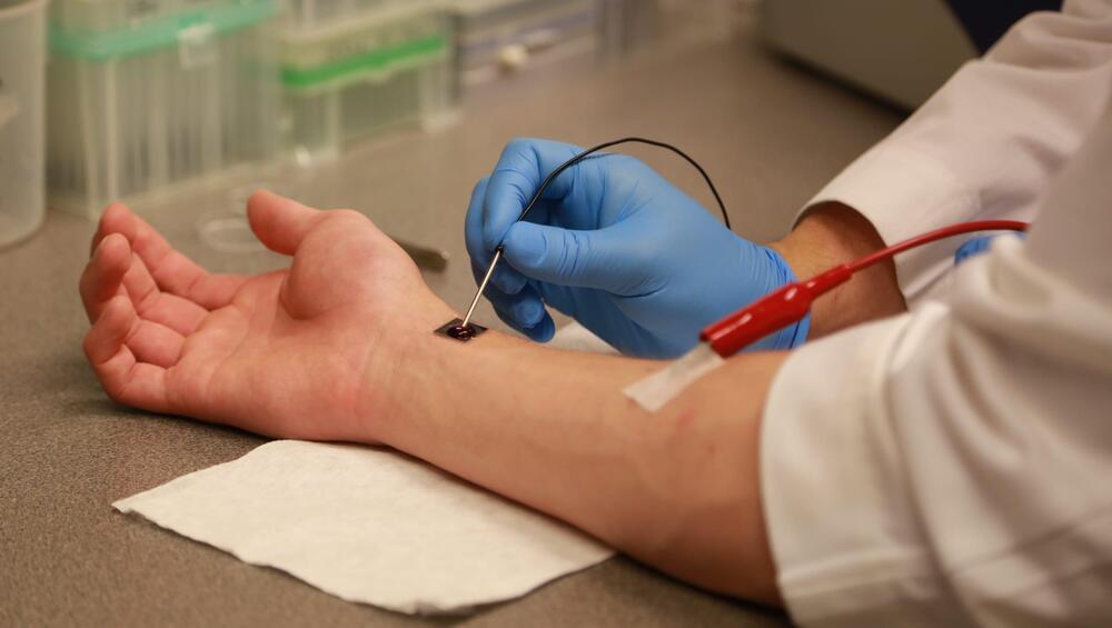
The first hallmark of aging is mutation. We can acquire mutations by being exposed to UV radiation or certain chemicals or through cell division. Cell divsion also leads to a second hallmark of aging (telomere attrition). Furthermore, our mitochondria start to work less as quality checks do not work properly anymore.
The hallmarks of aging are tightly linked to epigenetics. Epigenetics means that we have mechanisms (DNA methylation, histone modifications) which regulate the activity of genes. Epigenetics governs the development of embryonic stem cells into cells of our body but also impact aging. The loss of mitochondria for example is linked to dysfunctional epigenetic layers. As we age, at least three epigenetic modifications namely H4K16 acetylation, H3K4 trimethylation, or H4K20 trimethylation acumulate. The thing is that epigenetics is reversible… so can we also reverse aging?
Diets have been shown to slow down (and reverse aging to a small degree). Cells also show less damages in their DNA and we find higher levels of proteins which are found in “young cells. The activity of mitochondria is also increased if we undergo caloric restriction. Diets also impact the production of sirtuins which increase the lifespan and reverse aging. Different compounts (such as NMN and remodelin) have been shown to improve the epigenetic landscape which might have an effect on reversing aging. Exercise also might help to reverse aging as it helps to increase the activity of mitochondria. Meditation and having less stress also helps to increase the lengths of telomeres which might help to reverse aging. All in all studies suggests that some hallmarks of aging can be reversed so lets see where that goes!
0:00–0:46 Intro.
0:46–3:53 Hallmarks of Aging.
3:53–6:38 Epigenetics Controls Genes.
6:38–8:45 Reversing Aging: what is known.
8:45–11:25 Reversing Aging through Diets & Sports.
11:25–12:13 My Opinion.
References: