Wake Forest Institute for Regenerative Medicine (WFIRM) scientists working on CRISPR/Cas9-mediated gene editing technology have developed a method to increase efficiency of editing while minimizing DNA deletion sizes, a key step toward developing gene editing therapies to treat genetic diseases.
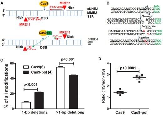
CRISPR (clustered regularly interspaced short palindromic repeats) technology is used to alter DNA sequences and modify gene function. CRISPR/Cas9 is an enzyme that is used like a pair of scissors to cut two strands of DNA at a specific location to add, remove or repair bits of DNA. By modifying gene function, scientists hope to treat genetic diseases by halting a diseased cell’s ability to continue replicating the defective DNA. CRISPR/Cas9 is the most versatile genetic manipulation available and has a wide range of potential applications. While CRISPR/Cas9 mainly generates short insertions or deletions at the target site, it may also make large DNA deletions around the specific target site. These large deletions cause safety concerns and may decrease functional editing efficiency.
The WFIRM team is looking for ways to reduce the chances of this happening. The research described in their recent paper, published recently in Nucleic Acids Research, sought to address the generation of unpredictable on-target long DNA deletions and find a way to guard against them, said lead author Baisong Lu, Ph.D., of WFIRM.