Toggle light / dark theme

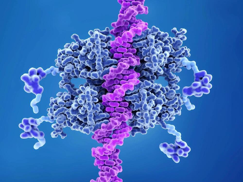
The label on RecA together with fluorescent markers on the DNA allows the researchers to follow every step of the process accurately; for example, they conclude that the whole repair is finished in 15 minutes, on average, and that the template is located in about nine. Using microscopy, Elf and his team investigate the fate of the break site and its homologous copy in real-time. They also find that the cell responds by rearranging RecA to form thin filaments that span the length of the cell.


How the cell can mend broken DNA

DNA, or deoxyribonucleic acid, is a molecule composed of two long strands of nucleotides that coil around each other to form a double helix. It is the hereditary material in humans and almost all other organisms that carries genetic instructions for development, functioning, growth, and reproduction. Nearly every cell in a person’s body has the same DNA. Most DNA is located in the cell nucleus (where it is called nuclear DNA), but a small amount of DNA can also be found in the mitochondria (where it is called mitochondrial DNA or mtDNA).

Watch the full documentary on Vimeo on demand: https://vimeo.com/ondemand/339083

The study of consciousness needs to be lifted out of the mysticism that has dominated it. Consciousness is not just a matter of philosophy or spirituality. It’s a matter of hard science. It’s a matter of understanding the brain and the mind — a pattern structure made out of information. It’s also a matter of engineering. If we can understand the functionality of the brain, its neural code, then we can build the same functionality into our computer systems. There’s no consensus on what produces consciousness, but everyone regardless of metaphysical views can agree what it is like to be conscious. Given that consciousness is subjectivity, what consciousness is like is what consciousness is.

‘Mind’ and ‘Consciousness’ are two different but somewhat overlapping terms related to the phenomenality of our experiential reality. Different species have a variety of their biological information processors which unsurprisingly results in qualia diversity. All species live in their own unique sensory universes. There is “something it is like to be” an organism. The human brain, our biological “wetware,” has a fractal structure on many genetic and abstract cognitive levels. Information is “modus operandi” of consciousness.

If we are to reason for the non-dual picture of the world then quantum physics is directly linked to consciousness. The human brain is a physical organ that transmits and interprets electrochemical signals. Its biochemistry is certainly governed by quantum physical laws, and consciousness — which is clearly related to the functioning of the brain — must therefore be related to the quantum physical processes going on within the brain and in the cosmos at large. Research has shown that consciousness is non-local, a scientific way of alluding to a connection within a higher dimensional order. Matter has also been shown to be non-local, which hints that matter might be an expression of consciousness, emerging from the ‘Unified Field’ — the quantum layer of pure potentiality — the code layer beneath all dimensions where time and space are information.

That fossil wasn’t enough to confirm Africa as our homeland. Since that discovery, paleoanthropologists have amassed many thousands of fossils, and the evidence over and over again has pointed to Africa as our place of origin. Genetic studies reinforce that story. African apes are indeed our closest living relatives, with chimpanzees more closely related to us than to gorillas. In fact, many scientists now include great apes in the hominid family, using the narrower term “hominin” to refer to humans and our extinct cousins.

In a field with a reputation for bitter feuds and rivalries, the notion of humankind’s African origins unifies human evolution researchers. “I think everybody agrees and understands that Africa was very pivotal in the evolution of our species,” says Charles Musiba, a paleoanthropologist at the University of Colorado Denver.

Paleoanthropologists have sketched a rough timeline of how that evolution played out. Sometime between 9 million and 6 million years ago, the first hominins evolved. Walking upright on two legs distinguished our ancestors from other apes; our ancestors also had smaller canine teeth, perhaps a sign of less aggression and a change in social interactions. Between about 3.5 million and 3 million years ago, humankind’s forerunners ventured beyond wooded areas. Africa was growing drier, and grasslands spread across the continent. Hominins were also crafting stone tools by this time. The human genus, Homo, arrived between 2.5 million and 2 million years ago, maybe earlier, with larger brains than their predecessors. By at least 2 million years ago, Homo members started traveling from Africa to Eurasia. By about 300,000 years ago, Homo sapiens, our species, emerged.

Dementia has many faces, and because of the wide range of ways in which it can develop and affect patients, it can be very challenging to treat. Now, however, using supercomputer analysis of big data, researchers from Japan were able to predict that a single protein is a key factor in the damage caused by two very common forms of dementia.

In a study published this month in Communications Biology, researchers from Tokyo Medical and Dental University (TMDU) have revealed that the HMGB1 is a key player in both frontotemporal lobar and Alzheimer , two of the most common causes of dementia.

Frontotemporal lobar degeneration can be caused by mutation of a variety of genes, which means that no one treatment will be right for all patients. However, there are some similarities between frontotemporal lobar degeneration and Alzheimer disease, which led the researchers at Tokyo Medical and Dental University (TMDU) to explore whether these two conditions cause damage to the brain in the same way.

The discriminatory power of the UVB variable is somewhat limited in this study, because UVB radiation is low at this time of the year, particularly at the high northern latitude of UK—larger effects might be observed if variation in UVB is greater. We only used ambient UVB, and did not capture individual behavioural differences that would determine the actual level of vitamin D synthesis in the skin, such as duration and time of day spent outside, clothing, etc. It is important to note that time of year is the strongest predictor of vitD-UVB. To avoid bias control dates were assigned to follow the same distribution as case dates, which might have led to artificially diminished differences in vitD-UVB between cases and controls, however analysis relating to hospitalisation and death are not affected by this. We also conducted an analysis of the genetically-predicted vitamin D and a number of state-of-the-art MR analyses. However, the main limitation is the lack of power. Given the small number of COVID-19 patients and the relatively small percentage of variance (4.2%) explained by vitamin D-related genetic variants, this MR study was not adequately powered to detect small causal effect and negative results should be interpreted with caution. Additionally, MR studies only consider linear effects between 25-OHD levels and COVID-19 risk, which do not capture what happens at the extremes of vitamin D deficiency. Therefore, it cannot rule out the possibility that seriously ill patients (due to an underlying pathology) with extremely low vitamin D levels could be predisposed to COVID-19 infection and increased COVID-19 severity and mortality. Furthermore, 25-OHD levels are the used biomarker of vitamin D status in the study population, nevertheless, they correlate poorly with the active form of vitamin D (1,25-OH2D), which exerts the effects of vitamin D on a cellular level. Thus, this study cannot exclude effects of 1,25-OH2D on COVID-19 risk.

Another limitation of this cohort relates to the fact that not all participants have been tested for present (or past) COVID-19 infection; consequentially, taking participants who were not tested as controls could be a potential source of bias, given that misclassification of controls might be substantial due to the presence of asymptomatic infected individuals, further driving our findings to the null. This is evident from the 1:2 ratio between outpatient vs. inpatient cases. It should be acknowledged that the COVID-19 cases in UK biobank have a high rate of hospitalisation due to the very limited and targeted testing at this stage of the pandemic in the UK, so this study reflects mainly those with more severe COVID-19 and gives less information about true infection risk, or risk of milder disease. In addition, we excluded individuals with a negative COVID-19 testing result from the controls due to the risk of those being false negatives. Although there is a risk of introducing selection bias, we believe that the risk of introducing misclassification bias if we included them in the analysis could be higher22,23. Additionally, given the presence of asymptomatic infected individuals, taking participants who were not tested as controls could also be another potential source of bias. Our study assessed the effect of genetically predicted vitamin D levels on COVID-19 risk while taking into consideration of ambient UVB radiation during the pandemic. We show an indication of an inverse association between genetically predicted vitamin D levels and severe COVID-19. Findings from our study are consistent with a recent randomised controlled trial (RCT) that found protective effect of vitamin D supplementation among those hospitalised with COVID-1924. However, other clinical trials did not show an effect. For instance, a randomised trial of 240 patients showed that supplementation with a single very large dose of 200,000 IU of vitamin D3 that increased serum vitamin D levels (21–44 ng/ml) was nonetheless ineffective in decreasing the length of hospital stay or any other clinical outcomes among hospitalized patients with severe COVID-1925. It has been estimated that one SD change in standardized natural-log transformed 25-OHD levels corresponds to a change in 25-OHD levels of 29.2 nmol/l in vitamin D insufficient individuals (serum 25-OHD levels 50 nmol/l), which is comparable to the 21.2 nmol/l mean increase in 25-OHD levels conferred by taking daily 400 IU of cholecalciferol, the amount of vitamin D most often found in vitamin D supplements26. This estimation has clinical implication on the dose of vitamin D supplement for disease prevention. Given the lack of highly effective therapies against COVID-19, it is important to remain open-minded to emerging results from rigorously conducted studies of vitamin D.

In conclusion, we found no significant associations between COVID-19 risk and measured 25-OHD levels after adjusted for covariates, but this finding is limited by the fact that the vitamin D levels were measured on average 11 years before the pandemic. Ambient UVB was strongly and inversely associated with COVID-19 hospitalization and death. The main MR analysis did not show that genetically-predicted vitamin D levels were causally associated with COVID-19 risk, although MR sensitivity analyses indicated a potential causal effect. Overall, the effect of vitamin D levels on the risk or severity of COVID-19 remains controversial, further studies are needed to validate vitamin D supplementation as a means of protecting against worsened COVID-19.

Because the herpesvirus sits in neurons forever, there is speculation it is connected to neurodegenerative diseases. The immune system requires inflammation to constantly fight off the virus, and neurons have some degree of damage because of this continuous immune response, according to Dr. Tibor Valyi-Nagy, professor of pathology, director of neuropathology at UIC and research collaborator on the study.


Summary: Researchers discovered mutations of the OPTN gene resulted in increased herpesvirus 1 growth in the brains of mice, leading to the death of local neurons. This resulted in accelerated neurodegeneration. OPTN deficiency was also associated with impairments in immune response. While these findings are specific to the HSV-1 virus, researchers believe the findings may apply to up to eight herpesvirus infections.

Source: University of Illinois at Chicago

A new study by researchers at University of Illinois Chicago suggests that when the protein optineurin, or OPTN, is present in cells it restricts the spread of HSV-1, the herpes simplex virus type 1.

As we age, our bones become thinner, we suffer fractures more often, and bone-diseases such as osteoporosis are more likely to occur. One responsible mechanism involves the impaired function of the bone-marrow stem cells, which are required for the maintenance of bone integrity. Researchers from the Max Planck Institute for Biology of Ageing and CECAD Cluster of Excellence for Ageing Research at the University of Cologne have now shown that the reduced stem cell function upon aging is due to changes in their epigenome. They were able to reverse these changes in isolated stem cells by adding acetate. This fountain of youth for the epigenome could become important for the treatment of diseases such as osteoporosis.

Aging Researchers have been looking at epigenetics as a cause of aging processes for some time. Epigenetics looks at changes in and chromosomes that do not alter the sequence of the genes themselves, but do affect their activity. One possibility is changes in proteins called histones, which package the DNA in our cells and thus control access to DNA. The Cologne research group of Peter Tessarz has now studied the epigenome of mesenchymal . These stem cells are found in marrow and can give rise to different types of cells such as cartilage, bone and fat cells.

This is an emerging therapeutic technique that works by targeting the underlying causes of a disease, rather than the symptoms it causes. It does this by targeting a particular gene, and preventing it from making the protein that it produces.


The United Kingdom’s NHS has very recently approved a new cholesterol-lowering jab which will be offered to 300,000 people over the next three years.

The drug – inclisiran – will be administered twice a year as an injection.

It will mainly be prescribed to patients who suffer with a genetic condition that leads to high cholesterol, those who have already suffered a heart attack or stroke, or those who haven’t responded well to other cholesterol-lowering drugs, such as statins.

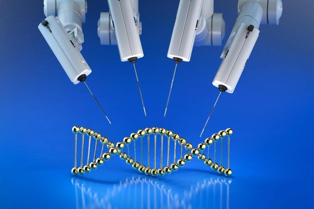
Within the last decade, scientists have adapted CRISPR systems from microbes into gene editing technology, a precise and programmable system for modifying DNA. Now, scientists at MIT’s McGovern Institute and the Broad Institute of MIT and Harvard have discovered a new class of programmable DNA modifying systems called OMEGAs (Obligate Mobile Element Guided Activity), which may naturally be involved in shuffling small bits of DNA throughout bacterial genomes.

These ancient DNA-cutting enzymes are guided to their targets by small pieces of RNA. While they originated in bacteria, they have now been engineered to work in human cells, suggesting they could be useful in the development of gene editing therapies, particularly as they are small (~30% the size of Cas9), making them easier to deliver to cells than bulkier enzymes. The discovery, reported in the journal Science, provides evidence that natural RNA-guided enzymes are among the most abundant proteins on earth, pointing toward a vast new area of biology that is poised to drive the next revolution in genome editing technology.

The research was led by McGovern investigator Feng Zhang, who is James and Patricia Poitras Professor of Neuroscience at MIT, a Howard Hughes Medical Institute investigator, and a core institute member of the Broad Institute. Zhang’s team has been exploring natural diversity in search of new molecular systems that can be rationally programmed.