Toggle light / dark theme

Summary: A new optogenetics-based technique allows researchers to control neuron excitability.

Source: MIT

Nearly 20 years ago, scientists developed ways to stimulate or silence neurons by shining light on them. This technique, known as optogenetics, allows researchers to discover the functions of specific neurons and how they communicate with other neurons to form circuits.

The world’s first artificial womb facility, EctoLife, will be able to grow 30,000 babies a year. It’s based on over 50 years of groundbreaking scientific research conducted by researchers worldwide.

Join me on:
- TikTok: https://www.tiktok.com/@hashem.alghaili.
- Instagram: https://www.instagram.com/hashem.alghaili/
- Facebook: https://www.facebook.com/ScienceNaturePage/
- LinkedIn: https://www.linkedin.com/in/hashem-al-ghaili-1b30679b/
- Twitter: https://twitter.com/HashemGhaili.

#EctoLife #ArtificialWomb #Genetics

Cancer, caused by abnormal overgrowth of cells, is the second-leading cause of death in the world. Researchers from the Salk Institute have zeroed in on specific mechanisms that activate oncogenes, which are altered genes that can cause normal cells to become cancer cells.

Cancer can be caused by , yet the impact of specific types such as structural variants that break and rejoin DNA, can vary widely. The findings, published in Nature on December 7, 2022, show that the activity of those mutations depends on the distance between a particular gene and the sequences that regulate the gene, as well as on the level of activity of the regulatory sequences involved.

This work advances the ability to predict and interpret which genetic mutations found in cancer genomes are causing the disease.

http://www.iBiology.org.

For millennia, humans have been harnessing #microbes to produce everything from breads, to cheeses, to alcohol. Now these tiny organisms have produced another powerful revolution — the gene editing tool CRISPR. Rodolphe Barrangou, Ph.D., was working at the food company Danisco, where he was trying to produce yogurt lines resistant to contamination. In a series of groundbreaking experiments, he helped uncover what CRISPR was, how it worked, and why it could be so transformative.

Speaker Biography:
Rodolphe Barrangou, Ph.D., studies beneficial microbes, focusing on the occurrence and diversity of lactic acid bacteria in fermented foods and as probiotics. Using functional genomics, he has focused on uncovering the genetic basis for health-promoting traits, including the ability to uptake and catabolize non-digestible carbohydrates. He spent 9 years at Danisco-DuPont, characterizing probiotics and starter cultures, and established the functional role of CRISPR-Cas as adaptive immune systems in bacteria. At NC State, he continues to study the molecular basis for their mechanism of action, as well as developing and applying CRISPR-based technologies for genotyping, building immunity and genome editing.

Producers: Sarah Goodwin, Rebecca Ellsworth.

A good night’s sleep can work wonders for both mind and body. But what is it that determines how much we need to sleep, and what can cause us to sleep more deeply?

In a new study, researchers from the University of Tsukuba have now provided some answers, revealing a signaling pathway within that regulates the length and depth of sleep.

“We examined in mice and how these affect their patterns of sleep,” says senior author of the study, Professor Hiromasa Funato. “We identified a mutation that led to the mice sleeping much longer and more deeply than usual.” The researchers found that this was caused by low levels of an enzyme called histone deacetylase 4 (HDAC4), which is known to suppress the expression of target genes.

CRISPR, the Nobel Prize-winning gene editing technology, is poised to have a profound impact on the fields of microbiology and medicine yet again.

A team led by CRISPR pioneer Jennifer Doudna and her longtime collaborator Jill Banfield has developed a clever tool to edit the genomes of bacteria-infecting viruses called bacteriophages using a rare form of CRISPR. The ability to easily engineer custom-designed —which has long eluded the —could help researchers control microbiomes without antibiotics or harsh chemicals, and treat dangerous drug-resistant infections. A paper describing the work was recently published in Nature Microbiology.

“Bacteriophages are some of the most abundant and diverse biological entities on Earth. Unlike prior approaches, this editing strategy works against the tremendous genetic diversity of bacteriophages,” said first author Benjamin Adler, a postdoctoral fellow in Doudna’s lab. “There are so many exciting directions here—discovery is literally at our fingertips.”

Imaging deep tissues with light is challenging. Visible light is often quickly absorbed and scattered by structures and molecules in the body, preventing researchers from seeing deeper than a millimeter within a tissue. If they do manage to probe further, substances like collagen or melanin often muddy the image, creating the equivalent of background noise through their natural fluorescence. As the authors explained, “Biological tissues have strong optical attenuation in the visible wavelength range (350–700 nm), due to the absorption of hemoglobin and melanin, as well as the tissue scattering, which fundamentally limits the imaging depth of high-resolution optical technologies.”

To wade out from these muddied waters, Yao and collaborator Vladislav Verkhusha, PhD, professor of genetics at Albert Einstein College of Medicine, developed a protein that absorbs and emits longer wavelengths of light in the near-infrared (NIR) spectrum. “Tissue is the most transparent in the 700‑1300 nm window of NIR light,” said Yao. “At those wavelengths, light can penetrate deeper into a tissue, and because there is less natural background fluorescence to filter out, we can take longer exposures and capture clearer images.”

Verkhusha and his lab used a process called directed molecular evolution to engineer their proteins, using photoreceptors normally found in bacteria as the basis for the structure. “The state-of-the-art NIR FPs were engineered from bacterial phytochrome photoreceptors (BphPs),” the team noted. “Applying rational design, we developed 17 kDa cyanobacteriochrome-based near-infrared (NIR-I) fluorescent protein, miRFP718nano.”

Breathing in common workplace dusts and fumes from agents such as vapors, gases, and solvents may heighten the risk of developing rheumatoid arthritis, suggests research published online in the Annals of the Rheumatic Diseases.

What’s more, such vapors, gases, and solvents seem to boost the detrimental impact of smoking and genetic susceptibility to the disease, the findings indicate.

Rheumatoid arthritis (RA) is a chronic autoimmune joint disorder characterized by painful and disabling inflammation. It affects up to 1% of the world’s population.

Modeling shows how genetic changes that don’t lead to changes in protein sequence can still alter protein function.

New modeling shows how synonymous mutations — those that change the DNA

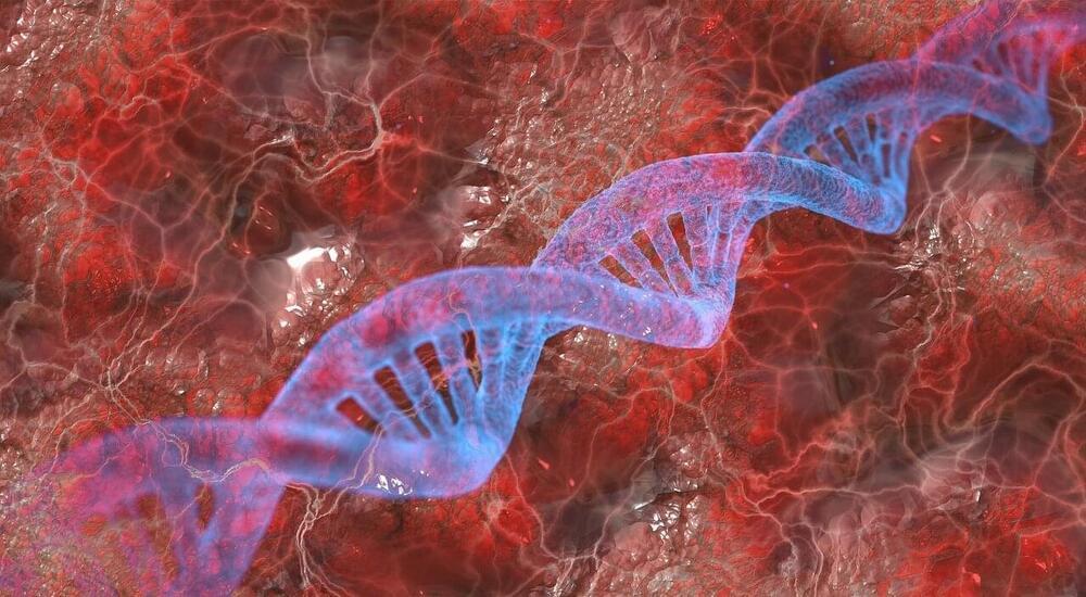
DNA, or deoxyribonucleic acid, is a molecule composed of two long strands of nucleotides that coil around each other to form a double helix. It is the hereditary material in humans and almost all other organisms that carries genetic instructions for development, functioning, growth, and reproduction. Nearly every cell in a person’s body has the same DNA. Most DNA is located in the cell nucleus (where it is called nuclear DNA), but a small amount of DNA can also be found in the mitochondria (where it is called mitochondrial DNA or mtDNA).

For the first cell to develop into an entire organism, genes, RNA molecules and proteins have to work together in a complex way. At first, this process is indirectly controlled by the mother. At a certain point in time, the protein GRIF-1 ensures that the offspring cut themselves off from this influence and start their own course of development. A research team from Martin Luther University Halle-Wittenberg (MLU) details how this process works in the journal Science Advances.

When a new organism starts to develop, the mother calls the shots. During fertilization, the and sperm fuse to form a single new cell. However, the course of , and thus how a new living being forms, is initially determined by the .

“Regardless of the organism, cell division is initially pre-programmed by the mother,” explains geneticist Professor Christian Eckmann from MLU. The mother’s cell provides a developmental starter set that includes the first proteins as well as the RNA molecules that serve as blueprints for further proteins. All this is necessary to jump start cell division and an organism’s development.