Toggle light / dark theme

Epigenetic signature for obesity found in study of twins

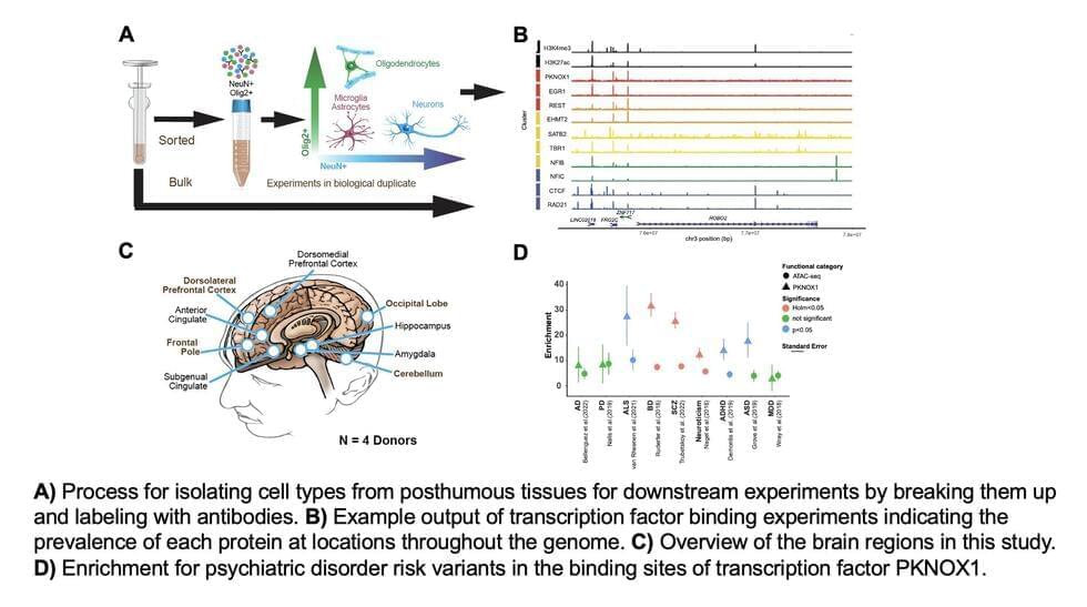
A susceptibility to gain weight may be written into molecular processes of human cells, a Washington State University study indicates.

The proof-of-concept study with a set of 22 found an epigenetic signature in buccal or cheek cells appearing only for the twins who were obese compared to their thinner siblings. With more research, the findings could lead to a simple cheek swab test for an obesity biomarker and enable earlier prevention methods for a condition that effects 50% of U.S. adults, the researchers said.

“Obesity appears to be more complex than simple consumption of food. Our work indicates there’s a susceptibility for this disease and molecular markers that are changing for it,” said Michael Skinner, a WSU professor of biology and corresponding author of the study published in the journal Epigenetics.

New treatment reverses epigenetic age of rats by 67.4%

Equivalent to an 80-year-old human reverting to the age of 26.


A groundbreaking study into anti-aging has reported significant rejuvenation effects using exosomes, tiny particles which can be extracted from biological fluids such as blood plasma.

Old and young rat. Image generated by DALL·E 3

In recent years, the prospect of being able to halt or even reverse aging has begun to seem less like science fiction and more like a scientific milestone that could emerge in the relatively near future.

Controlling organoids with light by combining spatial transcriptomics with optogenetics

They look like storm clouds that could fit on the head of a pin: Organoids are three-dimensional cell cultures that play a key role in medical and clinical research. This is thanks to their ability to replicate tissue structures and organ functions in the petri dish. Scientists can use organoids to understand how diseases occur, how organs develop, and how drugs work.

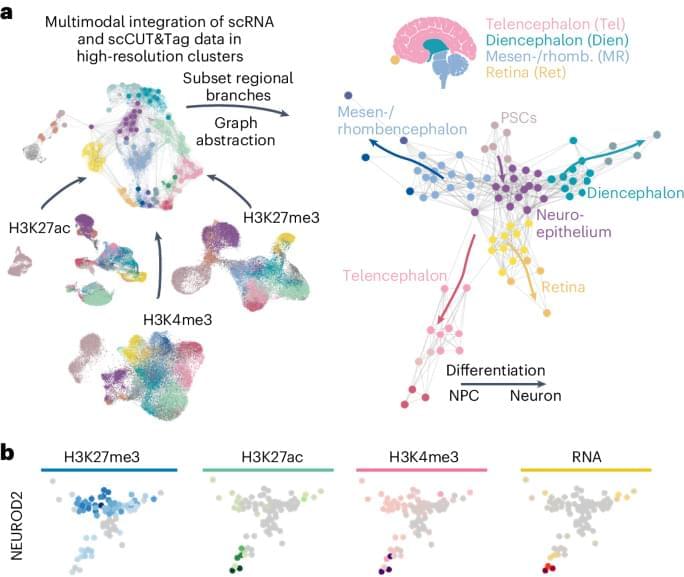
Single-cell technologies allow researchers to drill down to the molecular level of the cells. With spatial transcriptomics, they can observe which genes in the organoids are active and where over time.

The miniature organs are usually derived from . These are cells that haven’t differentiated at all, or only minimally. They can become any kind of cell, such as heart or kidney cells, , or neurons. To make stem cells differentiate, scientists “feed” them with growth factors and embed them in a nutrient solution.

NMN Ban? No Problem, Low Dose Niacin (60 mg/d) Increases NAD

Join us on Patreon! https://www.patreon.com/MichaelLustgartenPhD

Discount Links:
NAD+ Quantification: https://www.jinfiniti.com/intracellular-nad-test/
Use Code: ConquerAging At Checkout.

Epigenetic, Telomere Testing: https://trudiagnostic.com/?irclickid=U-s3Ii2r7xyIU-LSYLyQdQ6…M0&irgwc=1
Use Code: CONQUERAGING

At-Home Metabolomics: https://www.iollo.com?ref=michael-lustgarten.
Use Code: CONQUERAGING At Checkout.

Oral Microbiome: https://www.bristlehealth.com/?ref=michaellustgarten.
Enter Code: ConquerAging.

At-Home Blood Testing (SiPhox Health): https://getquantify.io/mlustgarten.

Re-Engineered Pseudoviruses for Precise and Robust 3D Mapping of Viral Infection

Engineered vesicular stomatitis virus (VSV) pseudotyping offers an essential method for exploring virus–cell interactions, particularly for viruses that require high biosafety levels. Although this approach has been employed effectively, the current methodologies for virus visualization and labeling can interfere with infectivity and lead to misinterpretation of results. In this study, we introduce an innovative approach combining genetic code expansion (GCE) and click chemistry with pseudotyped VSV to produce highly fluorescent and infectious pseudoviruses (clickVSVs). These clickVSVs enable robust and precise virus–cell interaction studies without compromising the biological function of the viral surface proteins. We evaluated this approach by generating VSVs bearing a unique chemical handle for click labeling and assessing the infectivity in relevant cell lines.

Study reveals how formaldehyde alters gene expression through epigenetics

Epigenetics, the chemical mechanisms that controls the activity of genes, allows our cells, tissues and organs to adapt to the changing circumstances of the environment around us. This advantage can become a drawback, though, as this epigenetic regulation can be more easily altered by toxins than the more stable genetic sequence of the DNA.

An article recently published at Science with the collaboration of the groups of Dr. Manel Esteller, Director of the Josep Carreras Leukaemia Research Institute (IJC-CERCA), ICREA Research Professor and Chairman of Genetics at the University of Barcelona, and Dr. Lucas Pontel, Ramon y Cajal Fellow also of the Josep Carreras Institute, demonstrates that the substance called formaldehyde, commonly present in various household and cosmetic products, in polluted air, and widely used in construction, is a powerful modifier of normal epigenetic patterns.

The publication is led by Dr. Christopher J. Chang, of the University of California Berkeley in the United States, whose research group is pioneer in the study of the effects of various chemical products on cell metabolism. The research has focused on investigating the effects of high concentrations of formaldehyde in the body, a substance already been associated with an increased risk of developing cancer (nasopharyngeal tumours and leukaemia), hepatic degeneration due to fatty liver (steatosis) and asthma. Dr. Esteller points out that this is relevant because “formaldehyde enters our body mainly during our breathing and, because it dissolves well in an aqueous medium, it ends up reaching all the cells of our body”.

Lipid Nanoparticle-Mediated Hit-and-Run Approaches Yield Efficient and Safe In Situ Gene Editing in Human Skin

Despite exciting advances in gene editing, the efficient delivery of genetic tools to extrahepatic tissues remains challenging. This holds particularly true for the skin, which poses a highly restrictive delivery barrier. In this study, we ran a head-to-head comparison between Cas9 mRNA or ribonucleoprotein (RNP)-loaded lipid nanoparticles (LNPs) to deliver gene editing tools into epidermal layers of human skin, aiming for in situ gene editing. We observed distinct LNP composition and cell-specific effects such as an extended presence of RNP in slow-cycling epithelial cells for up to 72 h. While obtaining similar gene editing rates using Cas9 RNP and mRNA with MC3-based LNPs (10–16%), mRNA-loaded LNPs proved to be more cytotoxic. Interestingly, ionizable lipids with a p Ka ∼ 7.1 yielded superior gene editing rates (55%–72%) in two-dimensional (2D) epithelial cells while no single guide RNA-dependent off-target effects were detectable. Unexpectedly, these high 2D editing efficacies did not translate to actual skin tissue where overall gene editing rates between 5%–12% were achieved after a single application and irrespective of the LNP composition. Finally, we successfully base-corrected a disease-causing mutation with an efficacy of ∼5% in autosomal recessive congenital ichthyosis patient cells, showcasing the potential of this strategy for the treatment of monogenic skin diseases. Taken together, this study demonstrates the feasibility of an in situ correction of disease-causing mutations in the skin that could provide effective treatment and potentially even a cure for rare, monogenic, and common skin diseases.

Running Large-Scale CRISPR Screens in Human Neurons

Identifying therapeutic targets for neurodegenerative conditions is often challenging due to the limited accessibility of reproducible, scalable in vitro cell models. Genome-level CRISPR screens are useful for these studies but performing screens that include the necessary replicates requires billions of cells. Human iPSC-derived cells can provide the needed scale, however, the complex process of directed differentiation is time-consuming, resource-intensive, and rarely feasible. Furthermore, delivering ribonucleases by transfection or transduction is inefficient in human iPSC-derived cells, especially delicate cell types like neurons. As a result, scientists often rely on immortalized cell lines, which do not accurately represent human biology or disease states, to run large-scale CRISPR screens.

In this GEN webinar, two experts will discuss solutions for running large-scale CRISPR screens to identify therapeutic targets for neurodegenerative diseases. They will present ioCRISPR-Ready Cells™: human iPSC-derived cells precision reprogrammed with opti-ox™, that constitutively express Cas9 nuclease, which are built for rapidly generating gene knockouts and CRISPR screens. During the webinar, you’ll learn about two peer-reviewed studies that performed large scale CRISPR knockout screens using opti-ox powered glutamatergic neurons with stable Cas9 expression. The first study demonstrates a loss-of-function genetic screen using a human druggable genome library. The second study investigated possible regulators of the RNA binding motif 3 protein, whose enhanced expression is highly neuroprotective both in vitro and in vivo.

Cancer Drug May Be Repurposed for Inflammatory Diseases

A cancer drug in the final stages of clinical trials may be able to help treat a range of inflammatory diseases including gout, heart failure, cardiomyopathy, and atrial fibrillation, according to scientists at the University of Cambridge.

Their findings are published in the Journal of Clinical Investigation in an article titled, “PLK1 inhibition dampens NLRP3 inflammasome-elicited response in inflammatory disease models.”

“Unabated activation of the NLR family pyrin domain–containing 3 (NLRP3) inflammasome is linked with the pathogenesis of various inflammatory disorders. Polo-like kinase 1 (PLK1) has been widely studied for its role in mitosis,” wrote the researchers. “Here, using both pharmacological and genetic approaches, we demonstrate that PLK1 promoted NLRP3 inflammasome activation at cell interphase. Using an unbiased proximity-dependent biotin identification (Bio-ID) screen for the PLK1 interactome in macrophages, we show an enhanced proximal association of NLRP3 with PLK1 upon NLRP3 inflammasome activation. We further confirmed the interaction between PLK1 and NLRP3 and identified the interacting domains.”

/* */