Toggle light / dark theme

Turning molecules into reliable electronic devices with a new fabrication platform

Molecules are among the smallest building blocks available for making next-generation devices. Their unique, customizable properties enable promising applications in emerging computing, sensing, optical and quantum technologies.

But integrating molecules into functional devices at scale remains a challenge. Traditional semiconductor manufacturing processes can damage small, fragile molecular materials. Now, MIT researchers have developed a scalable fabrication technique that incorporates delicate molecular materials into electronic devices on a chip without causing damage.

Their method extends the capabilities of standard semiconductor manufacturing processes to accommodate molecules. The researchers first prefabricate the device components using traditional processes. Then, they introduce the molecules and harness nanoscale surface forces to mechanically transform the fabricated device, which self-assembles without damaging the molecules.
Pixel patterns harness diffraction for faster, more accurate nanoscale 3D printing

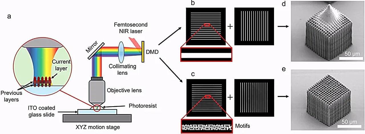
Researchers at the George W. Woodruff School of Mechanical Engineering have developed a new approach to nanoscale 3D printing that improves both speed and fidelity, overcoming a challenge that has limited the technology’s broader use in manufacturing.

Their work, led by Sourabh Saha, associate professor and Woodruff Faculty Fellow, and Harnjoo Kim, who conducted the research during his doctoral studies and later as a postdoctoral fellow in Saha’s lab, was recently published in the journal Nature Communications.

Nanoscale 3D printing allows researchers to create structures thousands of times smaller than the width of a human hair. These structures have potential applications in fields ranging from advanced computing and optics to biomedical devices and clean energy technologies. However, increasing printing speed often comes at the expense of quality.

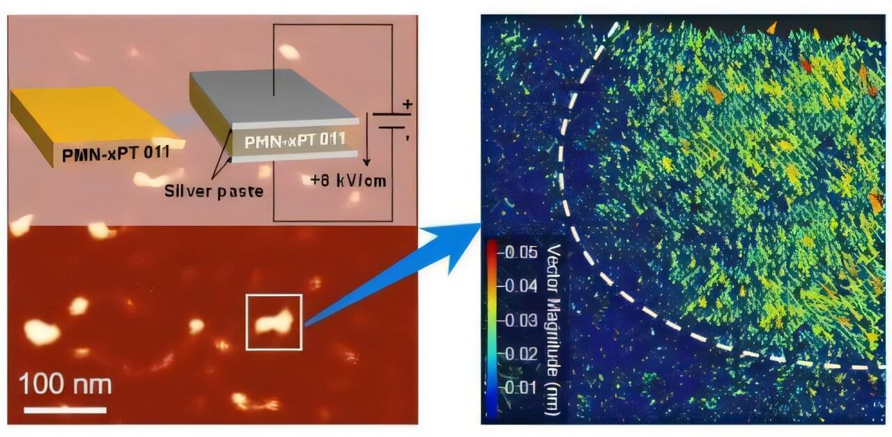
Light controls nanoscale ‘bubble’ domains in a ferroelectric crystal

Researchers at Flinders University have discovered an unexpected way light can control tiny electronic structures inside advanced materials, a development that could help pave the way for more energy-efficient memory devices, sensors and future computing technologies.

The new study, involving experts from around the world, explores electronic properties and optical science to uncover new energy and material capabilities.

“We discovered that light can control nanoscale ‘bubble’ domains (about the size of just a few billionths of a meter across) inside a special ferroelectric crystal,” says Dr. Pankaj Sharma, senior lecturer in experimental condensed matter physics at Flinders University.

New quantum encryption method prevents ciphertext from being cloned

Digital security currently relies on difficult equations to protect data. For example, when you use a credit card online, the information is locked inside a math problem that would take a modern computer thousands of years to solve. However, if someone builds a powerful enough computer, that security breaks.

To make systems safer, there is a major shift toward quantum security. This is where the unbreakable laws of quantum physics can be used to protect data instead.

Bringing it all into focus—computational microscope captures 25.2 billion pixels per second across a wide field of view

When designing microscopes, optical engineers have long faced a trade-off between speed, field of view and resolution. But improving one of these properties typically comes at the expense of another. Now, a UC Berkeley–led team of researchers has found a way to address this challenge, opening the door to new possibilities in the field of microscopy.

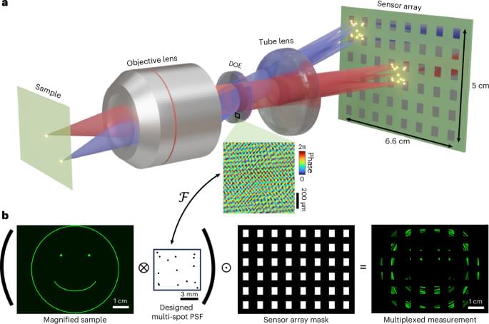
In a study published in Nature Photonics, researchers designed a computational microscope capable of capturing micron-scale resolution across multi-centimeter areas at faster-than-video rates. Using an array of 48 camera sensors and computational imaging “tricks,” their new microscope was able to capture videos at a rate of 25.2 billion pixels per second—an unprecedented combination of highly detailed magnification, wide field of view and high-speed imaging.

“This is really a breakthrough in the field of computational microscopy,” said Laura Waller, professor of electrical engineering and computer sciences and the study’s principal investigator. “With our microscope, we were able to achieve 3-micron resolution across 5 square centimeters at 120 frames per second, which isn’t possible with traditional ways. It’s the largest space-bandwidth time product of any practical microscope we know of.”

Decoding 3D chromatin architecture reveals distinct enhancer classes underlying hierarchical gene regulation in prostate cancer

The transcription process is controlled by non-coding regulatory elements, more than 70% of which are putative enhancers. These enhancers comprise over 600,000 regions and are marked by histone modifications. However, the mechanisms by which altered enhancers in cancer cooperate within the three-dimensional chromatin architecture to drive oncogenic programs remain poorly understood.

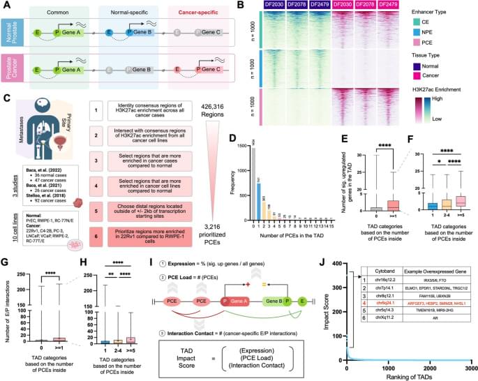
By integrating 201 H3K27ac ChIP-seq datasets from prostate, we identify 3,216 high-confidence prostate cancer-specific putative enhancers. Ultra-high-resolution chromatin interaction profiling by Region Capture Micro-C at a representative chr6q24.1 locus reveals that these enhancers form cancer-specific, highly nested interactions with promoters that coalesce into a multi-connected hub absent in normal prostate cells. CRISPR/Cas9 perturbations of these enhancers, examined one by one, distinguish enhancer classes within the hub. Deletion of a central enhancer collapses hub-wide enhancer activities and architecture, leading to the downregulation of target genes, impaired proliferation, and reduced clonogenic growth. In contrast, deletion of a redundant enhancer results in minimal transcriptional changes, as neighboring enhancers rescue cancer signaling through compensatory architectural rewiring that strengthens alternative enhancer-promoter interactions.

Largescale compressive microscopy via diffractive multiplexing across a sensor array Photonics

By combining an array of 48 sensors with a diffractive mask and compressive sensing, researchers developed a 25 gigapixel-per-second computational microscope that can image at micrometre resolution across multi-centimetre areas at 120 frames per second.

Two independent studies push semiconductor qubits towards practical scales

Semiconductor spin qubits are one of the most promising building blocks for future quantum computers, but turning them into a working, large-scale quantum computer has so far proven difficult. For now, two big questions remain open: how to connect qubits that aren’t sitting right next to each other, and how to control huge numbers of them without an unmanageable tangle of wiring.

Now, two independent studies published in Nature have each reported new experiments tackling these problems head-on.

Researchers Simulate Alzheimer’s Progression Across the Entire Brain

This hypothesis has led to a new “whole-brain” mathematical and computational model developed at the MOX Laboratory of the Department of Mathematics of Politecnico di Milano. The model is designed to describe in an integrated way the interaction between the spread of amyloid beta and the functioning of the cerebral vascular network. The aim is to provide a tool capable of simulating, on the scale of the whole organ, how small biological or vascular alterations can evolve over time and contribute to neurodegeneration. The model and the accompanying scientific study have been published in the prestigious scientific journal Computer Methods in Applied Mechanics and Engineering.

The model integrates two scales of analysis. On the one hand, it describes the dynamics of the production, transformation, diffusion and elimination of the healthy and pathological forms of amyloid beta. On the other, it describes blood flow through a “macroscopic” description of arteries, capillaries and veins, treating brain tissue as a porous medium perfused by blood vessels, through a macroscopic compartmental model. The two components are then connected to represent the possible mechanism of mutual reinforcement between protein accumulation and vascular dysfunction.

The simulations show a particularly relevant result: the brain can evolve into different states depending on the initial conditions. Small localised amounts of amyloid beta can be eliminated, allowing the system to return to a healthy state. Conversely, larger amounts can trigger a self-sustained spread of the pathology at brain scale.