A research team led by Prof. Sun Jianwei has achieved an advancement in organic synthesis and medicinal chemistry by developing an air-stable chiral phosphine-catalyzed enantioselective approach to synthesize enantioenriched S(IV)-stereogenic vinyl sulfinamides—an under-explored class of organosulfur compounds with promising antiviral activity.
The importance of chiral-at-sulfur compounds in drug discovery and organic synthesis is indisputable. More than a quarter of top-selling small molecule pharmaceuticals contain sulfur atoms, and chiral sulfinamides bearing S(IV) chirality are key building blocks for medicinal chemistry, asymmetric synthesis auxiliaries, and catalytic ligands. However, current methods to access enantioenriched sulfinamides rely on transition metal catalysis with organometallic nucleophiles, and efficient organocatalytic strategies have long remained unexplored, creating a critical gap in synthetic chemistry for this valuable chemical space.
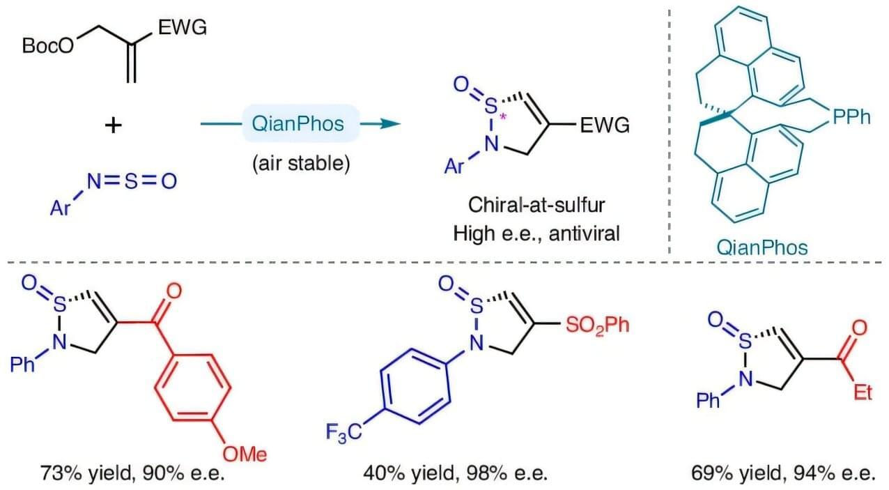
To address this challenge, Prof. Sun’s team published a study in Nature Chemistry detailing the design and synthesis of a novel C₂-symmetric chiral phosphine catalyst—QianPhos—derived from the SPHENOL chiral skeleton. This custom catalyst exhibits extraordinary air stability and structural rigidity, which enables highly chemo-, enantio-, and diastereoselective C−S bond formation via a [3+2] annulation between Morita–Baylis–Hillman (MBH) esters and sulfinylamines.


