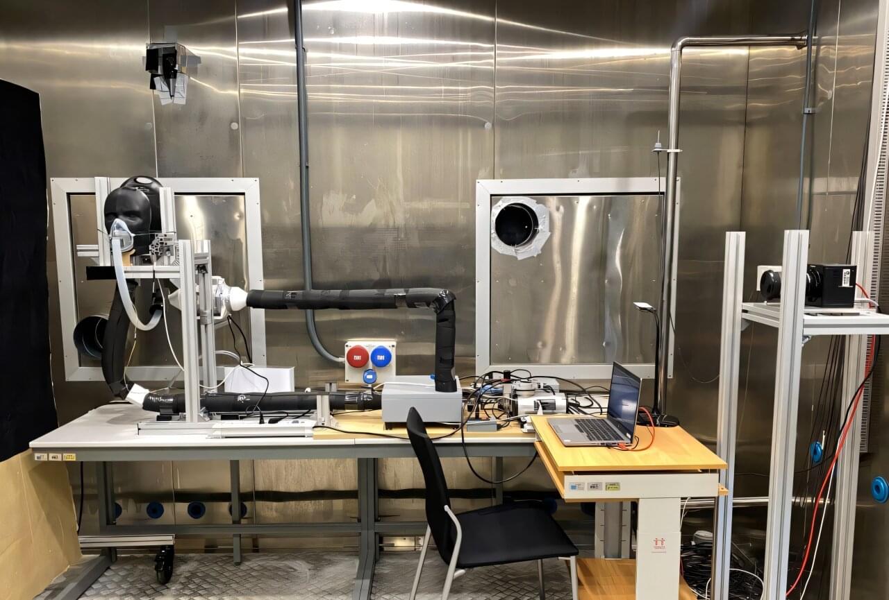
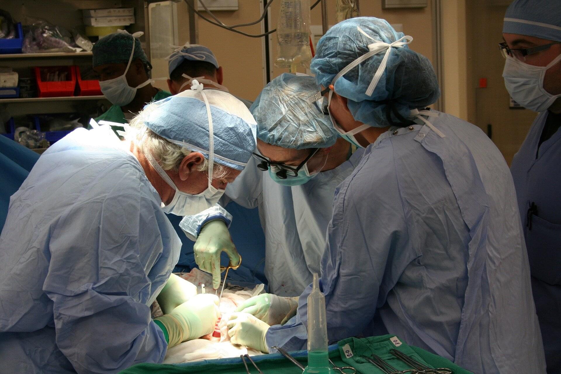
Toggle light / dark theme

Blood test can predict Alzheimer’s disease progression years before symptoms or brain scan changes

A study by investigators at Mass General Brigham has found that a blood test of plasma phosphorylated tau 217 (pTau217), an Alzheimer’s disease biomarker, can predict the progression of amyloid PET scan changes and cognitive decline in cognitively healthy older adults. The findings may help push back the clock to enable simpler, earlier disease prediction and indicate who may be at risk for cognitive decline. The results are published in Nature Communications.

“We used to think that PET scan detection was the earliest sign of Alzheimer’s disease progression, revealing amyloid accumulation in the brain 10 to 20 years before symptoms appear,” said lead author Hyun-Sik Yang, MD, a neurologist with Mass General Brigham Neuroscience Institute and an associate member of the Broad Institute of MIT and Harvard. “But now we are seeing that pTau217 can be detected years earlier, well before clear abnormalities appear on amyloid PET scans.”

Last year, the U.S. Food and Drug Administration cleared the first blood test for Alzheimer’s disease, paving the way for a cheaper, less invasive alternative to lumbar punctures and PET scans. The new study by Yang and colleagues adds important evidence about the predictive potential of these kinds of blood tests.

AI Model Can Help Cut Hospitalizations in Patients With Dialysis

AI models identified patients with end-stage kidney disease (ESKD) receiving hemodialysis who faced an imminent risk for hospital admission due to infections or fluid status abnormalities. When paired with nurse-led case reviews and targeted interventions, this strategy helped avert short-term admissions, demonstrating AI’s potential to guide timely, focused care.


AI-driven interventions reduce the odds of hospitalization within 7 days by 8% in patients with end-stage kidney disease receiving hemodialysis, according to a recent study.

Sibling Stem Cell Transplant Leads to Rare HIV Remission in ‘Oslo Patient’

After receiving a stem cell transplant from his brother, a 63-year-old Norwegian man known as the “Oslo patient” has become one of only a handful of people to see their HIV (human immunodeficiency virus) go into long-term remission.

While HIV can now be controlled with medication that stops the virus from replicating, the virus remains in the body, rebounding when the drugs are stopped. So case studies like this one are invaluable for researchers working towards a full cure.

The Oslo man was given a bone marrow stem cell transplant to treat a rare type of blood cancer. Discovering at the last minute that his brother carried a rare genetic mutation previously shown to resist HIV, researchers led by a team from Oslo University Hospital closely tracked the operation’s impact on the virus.

Proteomic insights into troponin elevation following COVID-19 infection

Background Raised cardiac troponin-I is a common finding in patients hospitalised with acute viral infections, including but not limited to COVID-19. This often occurs in the absence of overt myocardial injury presenting a challenge for interpretation. The mechanisms underlying troponin elevation are uncertain.

Methods The CISCO-19 (Cardiovascular Imaging in SARS-CoV-19) study (NCT04403607) is a prospective, multicentre cohort study, in which hospitalised PCR-confirmed COVID-19 participants (N=267) underwent multisystem evaluation at enrolment and at 28–60 days. The study incorporated plasma proteomics (SOMAscan V.4.1), cardiovascular MRI and clinical biomarkers. Of these, 211 had baseline plasma proteomic data and 185 completed follow-up sampling. Matched proteomic and imaging data were available for 155 participants (mean age: 55 years (SD 12); 43% female).

Results A high likelihood of myocarditis was identified in 13.2% (N=21/159) of participants. High-sensitivity troponin-I was modestly elevated at enrolment (median 3 ng/L; IQR 2–6; n=159). Among males (n=90), 9.3% had a high-sensitivity troponin that exceeded 34 ng/L. Among females (n=69), 4.5% exceeded 16 ng/L. Smooth muscle myosin light chain proteins were downregulated at follow-up (log2 fold change −0.12 to −0.6; all adjusted p0.02) and positively correlated with high-sensitivity troponin-I, but not N-terminal brain natriuretic peptide or cardiac MRI indices (n=155).

Association of Perinatal HIV Exposure and HIV Disease Severity With BP in Youth

RESEARCH ARTICLE: association of perinatal HIV exposure and HIV disease severity with BP in youth.


BACKGROUND: HIV infection is associated with cardiovascular events in adults. We compared mean blood pressure (BP) obtained at study visits between youth with/without perinatally acquired HIV infection and evaluated whether HIV disease severity was associated with BP. METHODS: BP was compared between participants with/without HIV in the Adolescent Master Protocol of the Pediatric HIV/AIDS Cohort Study. Marginal repeated measures analyses using generalized estimating equations evaluated the association of HIV disease severity with BP index (mean BP/95th percentile BP) and abnormal BP. RESULTS: 447 youth with HIV and 226 youth without HIV were included. Youth with HIV were more often Black non-Hispanic (66% versus 54%), had greater household income (54% versus 35%), and lower measures of adiposity than those without.

Integrative approaches to aging: Mechanisms, antiaging strategies, and emerging biomedical interventions

This imbalance results in dermal thinning, wrinkle formation, and loss of skin elasticity. Both intrinsic aging (chronological) and extrinsic aging (photoaging) contribute to collagen depletion. Studies have shown that UV-induced ROS accelerate collagen breakdown and inhibit new collagen synthesis, exacerbating visible signs of aging. [20]

Collagen is vital for skin firmness and elasticity. Aging, both intrinsic and extrinsic, leads to reduced collagen production and increased enzymatic degradation. Antiaging interventions such as retinoids, marine peptides, and nanoformulations aim to restore collagen levels and improve skin structure.

Understanding these cellular and molecular mechanisms provides the foundation for developing targeted antiaging interventions, ranging from holistic lifestyle modifications to advanced biomedical therapies.

HarmonyGNN boosts graph AI accuracy on four tough benchmarks by up to 9.6%

Researchers have demonstrated a new training technique that significantly improves the accuracy of graph neural networks (GNNs)—AI systems used in applications from drug discovery to weather forecasting. GNNs are AI systems designed to perform tasks where the input data is presented in the form of graphs. Graphs, in this context, refer largely to data structures where data points (called nodes) are connected by lines (called edges). The edges indicate some sort of relationship between the nodes. Edges can be used to connect nodes that are similar (called homophily)—but can also connect nodes that are dissimilar (called heterophily).

For example, in a graph of a neural system there would be edges between nodes representing two neurons that enhance each other, but there would also be edges between nodes that suppress each other.

Because graphs can be used to represent everything from social networks to molecular structure, GNNS are able to capture complex relationships better than many other types of AI systems.

Minimally Invasive Ablation Can Treat Small Kidney Tumors

Among patients with T1a renal cell carcinoma (T1a RCC), ablation and surgical resection showed comparable risks for tumor progression. However, ablation was associated with higher rates of local recurrence but fewer complications and shorter hospital stays than resection or nephrectomy.


“Follow-up data revealed that most local recurrences in patients who underwent ablation were successfully treated with additional ablation or surgery,” the authors wrote.

“[T]his study suggests ablation as a less invasive alternative to surgery for patients with T1a RCC, resulting in a similar high level of oncologic control,” they added.

This study was led by Johanne Ahrenfeldt, PhD, MScEng, Aarhus University Hospital, Denmark. It was published online in Radiology.

APOE4, the Alzheimer’s risk gene, silently undermines bone quality in women

Scientists at the Buck Institute for Research on Aging, along with collaborators at UC San Francisco, have discovered that APOE4, the most common genetic risk factor for Alzheimer’s disease, causes bone quality deficits specifically in female mice, through a mechanism that is invisible to standard imaging and can emerge as early as midlife. The findings, published in Advanced Science, reveal an unexpected biological link between Alzheimer’s risk and skeletal health, and identify a new molecular pathway that could one day inform earlier diagnosis of cognitive decline or guide treatment for bone quality loss in women who carry the APOE4 gene.

“What makes this finding so striking is that bone quality is being compromised at a molecular level that a standard bone scan simply will not catch,” says Buck professor Birgit Schilling, Ph.D., a senior author of the study. “APOE4 is quietly disrupting the very cells responsible for keeping bone strong, and it is doing this specifically in females, which mirrors what we see with Alzheimer’s disease risk.”

Physicians have long observed that people with Alzheimer’s disease suffer bone fractures at higher rates, and that a diagnosis of osteoporosis in women is actually the earliest known predictor of Alzheimer’s. But the underlying mechanism connecting brain and bone health has remained elusive.

/* */