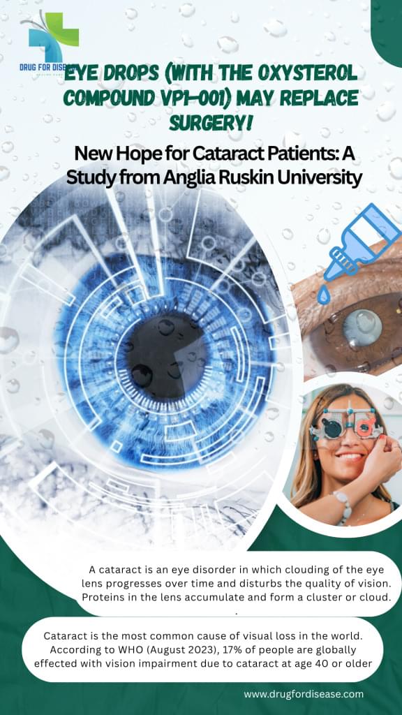
A study from Anglia Ruskin University showed that eye drops with oxysterol compound (VP1-001) may treat cataracts without surgery. The study’s results were published on May 2nd, 2022, in the peer-reviewed journal Investigative Ophthalmology and Visual Science. A cataract is an eye disorder in which clouding of the eye lens worsens over time and disrupts the quality of vision. A cataract is a disorder in which the proteins in the lens accumulate & make a cluster or cloud. This cloud scatters light and significantly limits its transmission to the retina. Cataract is the most common cause of visual loss in the world. According to the report of WHO (10 August 2023), 17% of people are globally affected with vision impairment due to cataracts at age 40 or older. At present, cataract surgery is the only way for the treatment of cataracts. In this surgical procedure the hazy lens is replaced with an artificial intraocular lens. Including economical burdens, cataract surgery has many complications like inflammation, xerophthalmia macular oedema, and posterior capsular opacificationHowever, a few scientists at Anglia Ruskin University under the supervision of Prof. Barbara, Deputy Dean in the Faculty of Health, Education, Medicine, and Social Care, conducted optical tests on an oxysterol compound that is considered an anti-cataract drug. They prepared new eye drops that could get rid of cataracts without surgery. VP1-001 is a chemical that is in these drops. It works by repairing the protein clumps in the eye lens that make it cloudy. A single drop increased the lens’s clarity and focusing capacity when tested on mice with cataracts. The study’s results were published on May 2nd, 2022, in the peer-reviewed journal Investigative Ophthalmology and Visual Science. Outcomes of optical tests with eye drops The outcomes were good: 61% of the eyes that were treated were better able to focus due to the improvement in the refractive index profiles 46% of them became clearer and more transparent. Results: This is a huge advance since it means that some cataracts might be able to be cured with drugs instead of surgery. But the drops didn’t work on all kinds of cataracts, so additional research is needed to find therapies that work for everyone. Moreover, these drops aren’t available for individuals now. More research is needed These drops aren’t available for individuals now, but this is a big step forward, especially in countries where eye surgery is hard to get. This study is a hope towards non-surgical treatment of cataracts with the oxysterol compound (VP1-001). Cataract surgery is a safe and effective solution, as it completely replaces the clouded lens with an artificial one. The oxysterol compound (VP1-001) is still in the preclinical stage and not approved for human use. It’s under clinical trials. Moreover, safety testing will be confirmed before public availability. 📌 Published in: Investigative Ophthalmology and Visual Science (May 2, 2022) by Anglia Ruskin University Frequently asked questions 1. What are the types of cataracts? Caract can be classified by many wayss; however, it can be categorised into three types, on the basis of cloud location/formation, that are Posterior subcapsular cataract Age-related cataracts. Cortical cataracts. Nuclear Cataracts. 2. What are the risk factors of cataracts? Age is the major factor of cataracts, especially at 40 or above. other factors include diabetes, smoking, obesity, hypersensitivity, excessive exposure to sunlight, eye injury or inflammation, etc. 3. Are there any eye drops or drugs to treat cataracts without surgery? As of now (2025), there is no approved eye drop or drug that can treat or reverse cataracts without surgery, but research at Anglia Ruskin University showed that the experimental drug VP1-001 (oxysterol-based eye drops) works as an anti-cataract in animal trials. 4. What are the complications of cataract surgery? Many complications, like inflammation, xerophthalmia, and macular oedema, can occur as post-surgical conditions. 5. What is the oxysterol compound VP1-001? Oxysterol compound VP1-001, also known as compound 29, has shown a significant effect on the treatment of cataracts in a trial on mice. It reduces the opaqueness of the lens that may occur due to risk factors like ageing & mutation. Premium SEO Backlinks


