A new study probing quantum phenomena in neurons as they transmit messages in the brain could provide fresh insight into how our brains function.
In this project, described in the Computational and Structural Biotechnology Journal, theoretical physicist Partha Ghose from the Tagore Centre for Natural Sciences and Philosophy in India, together with theoretical neuroscientist Dimitris Pinotsis from City St George’s, University of London and the MillerLab of MIT, proved that established equations describing the classical physics of brain responses are mathematically equivalent to equations describing quantum mechanics. Ghose and Pinotsis then derived a Schrödinger-like equation specifically for neurons.
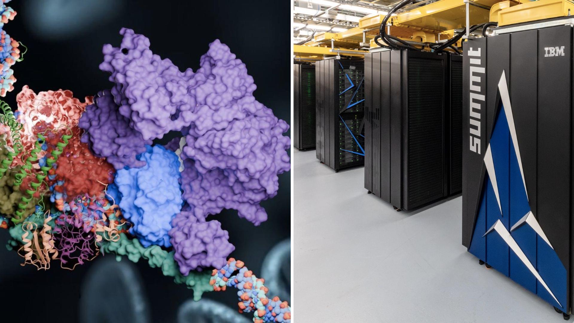
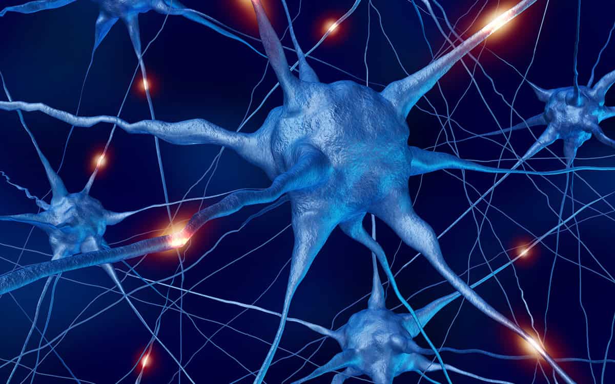
Our brains process information via a vast network containing many millions of neurons, which can each send and receive chemical and electrical signals. Information is transmitted by nerve impulses that pass from one neuron to the next, thanks to a flow of ions across the neuron’s cell membrane. This results in an experimentally detectable change in electrical potential difference across the membrane known as the “action potential” or “spike”