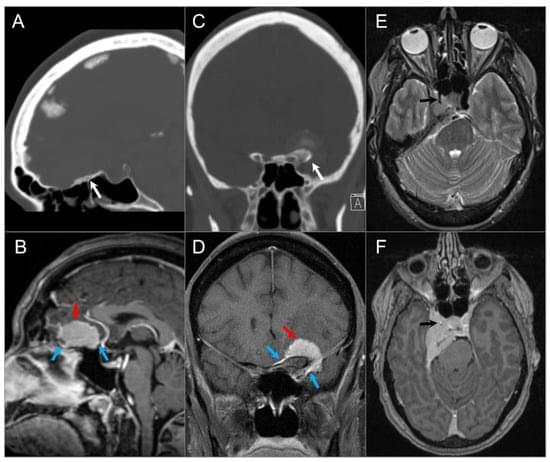
Toggle light / dark theme

Exposure to nanoplastics could induce spread of Alzheimer’s disease from the brain to other organs

A new preclinical study has found exposure to nanoplastics may contribute to the rapid progression of Alzheimer’s disease and subsequent spread from the brain to other key organs such as the liver, heart and gut.

The research, “Cerebral to Systemic Representations of Alzheimer’s Pathogenesis Stimulated by Polystyrene Nanoplastics,” is published in the journal Environment & Health.

The study, co-led by Monash University and South China University of Technology, investigated how environmental-level polystyrene exposure influences the progression of Alzheimer’s disease from the brain to other parts of the body. Studies in mice revealed that nanoplastic-induced neurological damage is not confined within the brain, but expands systemically through the gut–liver–brain axis.

Detecting early-stage tumors with a blood sample

Current methods for cancer diagnosis are based on identifying biomarkers — molecules that reveal a particular state or process in the body – produced by the tumor or associated proteins. Not surprisingly, these markers are more abundant once the tumor has already developed significantly. And the more advanced the tumor, the more difficult it is to find effective treatment options.

Now, a research team has developed a test that can detect early-stage solid tumors with just a blood sample. In addition, the test also provides information relevant to the choice of treatment.

To achieve this early detection, the team focused the test not on the markers produced by the tumor, but on the body’s defensive reaction to the cancer. Since the 19th century it has been known that the emergence of cancer cells causes changes in the immune system, and it was also known that these changes are more intense in cancer’s earliest stages. But they had never been used for diagnosis. The new study focuses on them, specifically on the changes in blood proteins derived from cancer’s disruption of the immune system.

But this approach posed a problem to the team: human blood contains more than 5,000 proteins, which makes it extremely difficult to analyze. So they used bioinformatics analysis and narrowed the scope of the study to five amino acids: lysine, tryptophan, tyrosine, cysteine and cysteine not bound to disulphide bonds.

They then subjected the sample to reactions that emit fluorescence when light is applied to them — fluorogenic reactions — and revealed the exact concentration of each of these amino acids in the plasma. Using the artificial intelligence tool machine learning, they identified patterns in these concentrations that could be translated into diagnostic signals.

As they explain in the published article, they applied this technique to samples from 170 patients and were able to identify 78% of cancers with a 0% false positive rate.

Human CLOCK gene enhances brain connectivity and mental flexibility in mice, study finds

Clock genes are a set of genes known to contribute to the regulation of the human body’s internal 24-hour cycle, also known as the circadian rhythm. One of these genes is the so-called CLOCK gene, a protein that regulates the activity of other genes, contributing to recurrent patterns of sleep and wakefulness.

Past findings suggest that this gene is also expressed in the neocortex, a brain region that supports important cognitive abilities, including reasoning, decision-making and the processing of language. However, the gene’s possible contribution to these specific brain functions remains poorly understood.

Researchers at UT Southwestern Medical Center recently carried out a study on genetically modified mice aimed at better understanding how the expression of the CLOCK gene in the human neocortex influences cognitive functions. Their findings, published in Nature Neuroscience, suggest that the gene plays a role in the formation of connections between neurons, which in turn influence mental and behavioral flexibility.

When does the body really start aging? The answer may surprise you

When does aging really shift into overdrive? A new study suggests it may be sooner than you think.

Scientists at the Chinese Academy of Sciences studied proteins in tissue taken from about 70 people ages 14 to 68, according to the study published July 25 in the journal Cell.

These proteins give scientists a window of when the aging process may begin on a cellular level, said Dr. Thomas Blackwell, associate dean for graduate medical education and professor of medicine at the University of Texas Medical Branch.

New research unveils vast influence of B vitamins on health and disease

Eight essential nutrients make up the suite of B vitamins also known as the B complex. Researchers from Tufts University and elsewhere have revealed that these B vitamins influence a vast spectrum of human health and disease, including cognitive function, cardiovascular health, gastric bypass recovery, neural tube defects, and even cancer.

“It’s hard to study the B vitamins in isolation,” says gastroenterologist Joel Mason, senior scientist at the Jean Mayer USDA Human Nutrition Research Center on Aging (HNRCA) and professor at the Gerald J. and Dorothy R. Friedman School of Nutrition Science and Policy and Tufts University School of Medicine. “Four of these B-vitamins cooperate as co-factors in many critical activities in cells in what we call ‘one carbon metabolism’.”

One carbon metabolism is a series of pathways that allow for the transfer of single-carbon units to cells for essential processes such as DNA synthesis, amino acid metabolism, and more. It’s their role in all these crucial biological functions that make the B vitamins so important-and so challenging to tease out how they contribute positively and, perhaps negatively, to human health.

MIT engineers uncover a surprising reason why tissues are flexible or rigid

Water makes up around 60 percent of the human body. More than half of this water sloshes around inside the cells that make up organs and tissues. Much of the remaining water flows in the nooks and crannies between cells, much like seawater between grains of sand.

Now, MIT engineers have found that this “intercellular” fluid plays a major role in how tissues respond when squeezed, pressed, or physically deformed. Their findings could help scientists understand how cells, tissues, and organs physically adapt to conditions such as aging, cancer, diabetes, and certain neuromuscular diseases.

In a paper appearing today in Nature Physics, the researchers show that when a tissue is pressed or squeezed, it is more compliant and relaxes more quickly when the fluid between its cells flows easily. When the cells are packed together and there is less room for intercellular flow, the tissue as a whole is stiffer and resists being pressed or squeezed.

New carbon material sharpens proton beams, potentially boosting cancer treatment precision

Researchers from the National University of Singapore (NUS) have developed a groundbreaking carbon membrane that could revolutionise proton therapy for cancer patients, and advance technologies in medicine and other areas such as energy devices and flexible electronics.

The new carbon material which is just a single atom thick shows incredible promise in enabling high-precision proton beams. Such beams are key to safer and more accurate proton therapy for cancer treatment. The new material, called the ultra-clean monolayer amorphous carbon (UC-MAC), could outperform best in class materials like graphene or commercial carbon films.

The research was led by Associate Professor Lu Jiong and his team from the NUS Department of Chemistry, in collaboration with international partners.

New Technique Uses Focused Sound Waves and Holograms to Control Brain Circuits

NEW YORK, Aug. 5, 2025 /PRNewswire/ — A new study provides the first visual evidence showing that brain circuits in living animals can be activated by ultrasound waves projected into specific patterns (holograms).

Led by scientists at NYU Langone Health and at the University of Zurich and ETH Zurich in Switzerland, the study describes a system that combines sources of ultrasound waves and a fiber scope connected to a camera to visualize in study mice brain targets that are directly activated by the sound. This lays the groundwork, the study authors say, for a new way to treat neurological diseases and mental health disorders from outside of the body.

Already, there are applications approved by the Food and Drug Administration and designed to reduce tremor symptoms seen in Parkinson’s disease, using intense sound waves to kill brain cells called neurons within neural pathways linked to tremors. Rather than kill neurons, the lower-intensity ultrasound waves used in the current work can temporarily activate them, the researchers say. The resulting effects can be widespread as neurons relay messages to other neurons within their circuits and between interconnected neuronal circuits.

Metabolic signals in neurons determine whether axons degrade or resist neurodegeneration, study finds

Unlike most cells in the human body, neurons—the functional cells of our nervous system—cannot typically replace themselves with healthy copies after being damaged.

Rather, after an injury from something like a stroke, concussion or neurodegenerative disease, neurons and their axons, fiber-like projections that relay , are far more likely to degrade than regenerate.

But new research from the University of Michigan opens new ways to think about neurodegeneration that could help protect patients against that degradation and neurological decline in the future.

Stem cells created from ALS patients point to potential new target for treatment

Amyotrophic lateral sclerosis (ALS), known as Lou Gehrig’s disease, is an incurable neurological disorder affecting motor neurons—nerve cells in the brain and spinal cord that control voluntary muscle movement and breathing.

Many ALS , including those testing promising drugs, have fallen short of expectations—often because the extent of the disease can vary, and patients don’t respond the same way to medications.

But a new study led by scientists at Case Western Reserve University used created from ALS patients to target a specific gene as a kind of shut-off valve for what stresses —and it worked.

/* */