A look inside Mindstate Design Labs’ effort to design drugs that reliably produce specific states of consciousness.

An unusual public health policy in Wales may have produced the strongest evidence yet that a vaccine can reduce the risk of dementia. In a new study led by Stanford Medicine, researchers analyzing the health records of Welsh older adults discovered that those who received the shingles vaccine were 20% less likely to develop dementia over the next seven years than those who did not receive the vaccine.
The remarkable findings, published in Nature, support an emerging theory that viruses that affect the nervous system can increase the risk of dementia. If further confirmed, the new findings suggest that a preventive intervention for dementia is already close at hand.

A revolution is underway in gene editing—and at its forefront is David Liu, an American molecular biologist whose pioneering work is rewriting the building blocks of life with unprecedented precision.
A professor at the Broad Institute of MIT and Harvard, Liu was awarded a Breakthrough Prize in Life Sciences on Saturday for developing two transformative technologies: one already improving the lives of patients with severe genetic diseases, the other poised to reshape medicine in the years ahead.
He spoke with AFP ahead of the Los Angeles ceremony for the prestigious Silicon Valley-founded award.
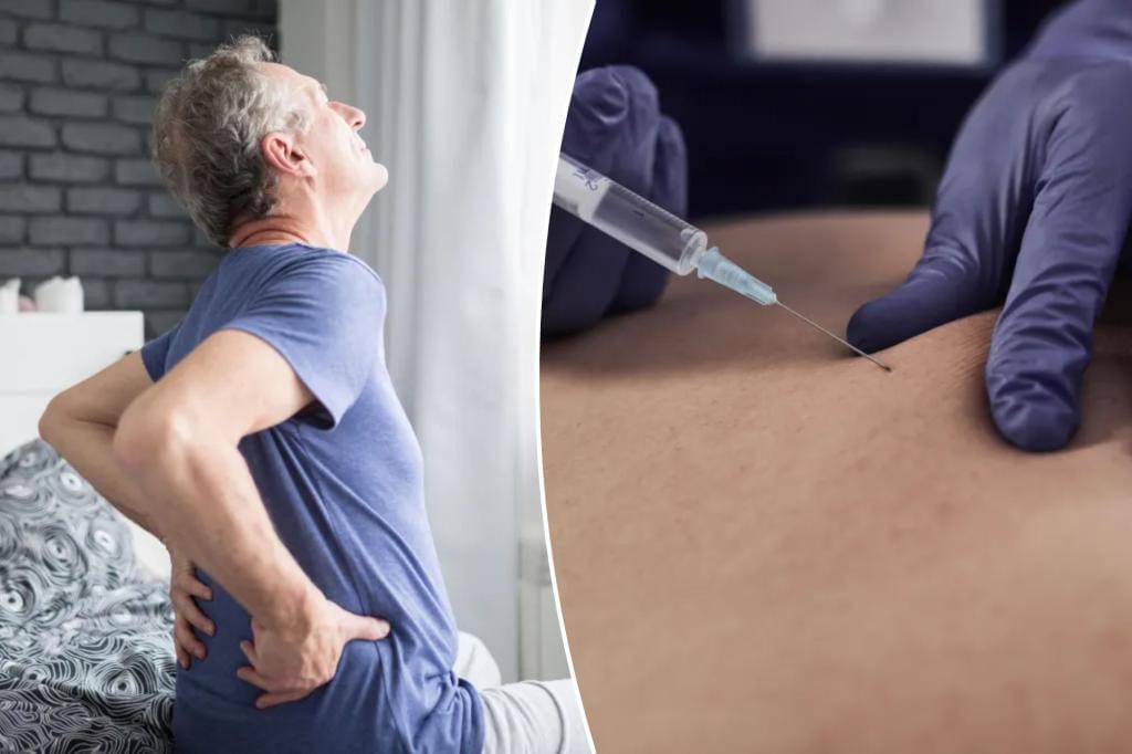
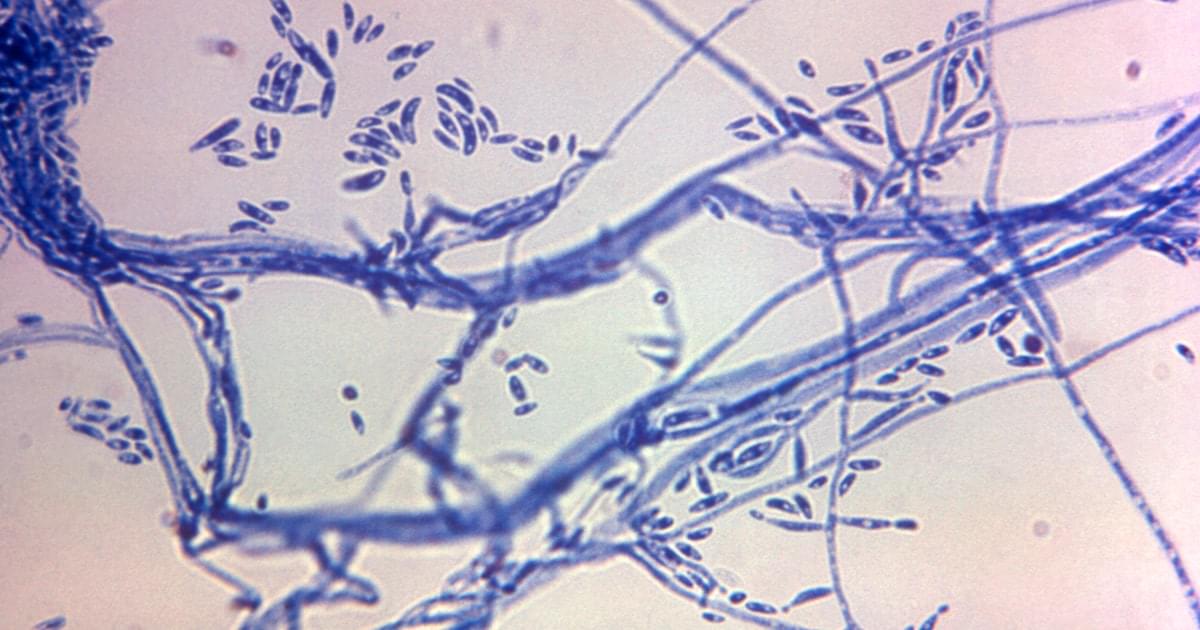
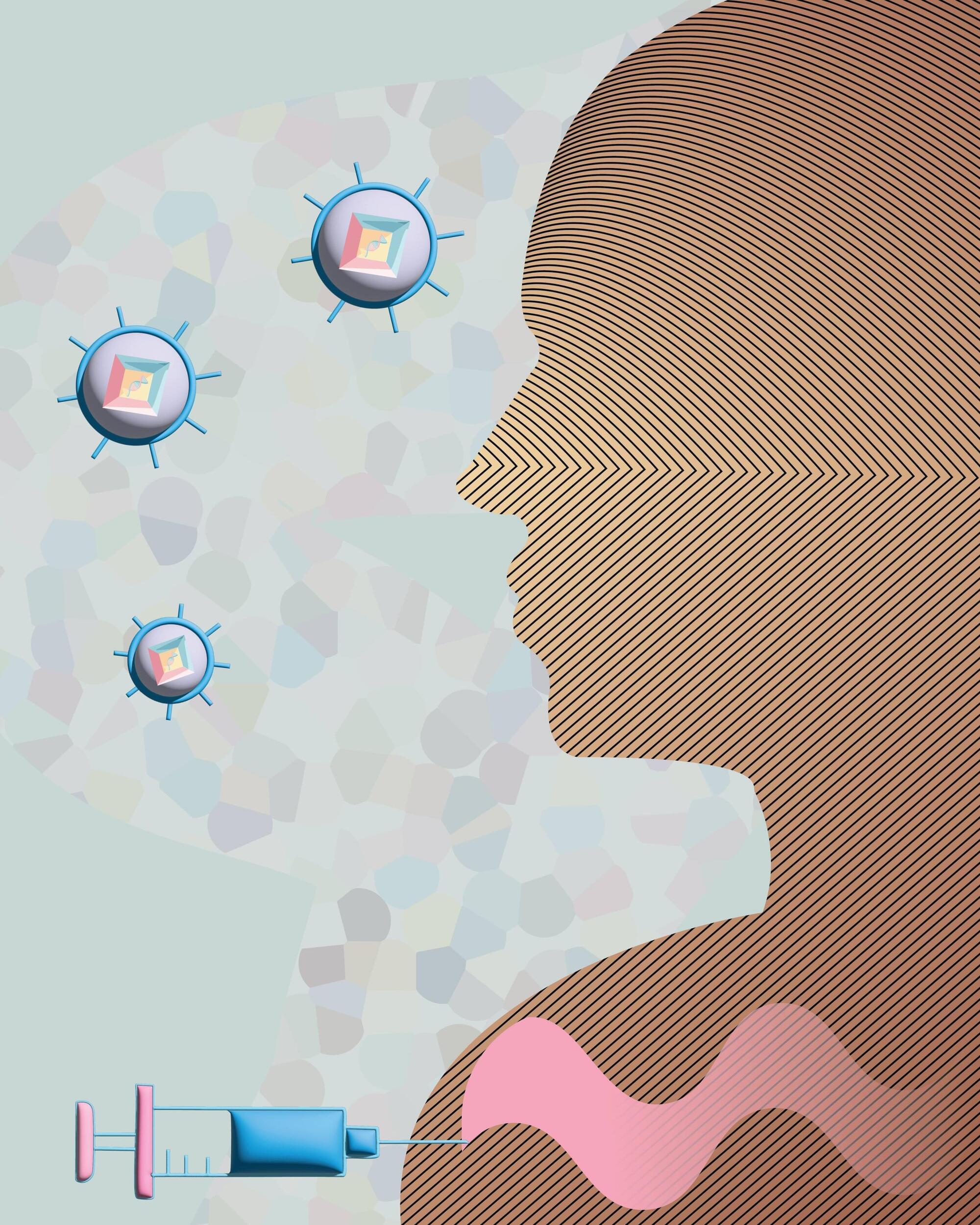
Chronic lower back pain is one of the top complaints that sends Americans to their doctors — and it’s a leading cause of missed workdays and disability claims.
While slipped discs, arthritis and spinal problems are often blamed, for some, the real culprit is an infection. Now, there’s hope on the horizon for these patients.
Early clinical trial results indicate a new antibiotic drug could treat — or even cure — the infection. Experts are hailing it as a “massive gamechanger” with the potential to drastically improve the quality of life for those suffering from chronic lower back pain.
EPFL’s Blue Brain Project deployed its powerful brain simulation technology and expertise in cellular and molecular biology to try and answer this question.

The clinical treatment of profound oxygen deprivation (hypoxemia) is time sensitive and requires skill and specialized equipment. When the airways or lungs become incapacitated (e.g., due to airway obstruction or lung injury), resuscitation is ineffective until oxygenation is restored. Many critically ill patients suffer organ dysfunction, cardiac arrest, or death within minutes. In this work, we describe a polymeric microparticle-based oxygen delivery technology capable of rapidly administering large volumes of oxygen gas through an intravenous line.
ABSTRACT: A continuous supply of oxygen to tissues is vital to life and interruptions in its delivery are poorly tolerated. The treatment of low-blood oxygen tensions requires restoration of functional airways and lungs. Unfortunately, severe oxygen deprivation carries a high mortality rate and can make otherwise-survivable illnesses unsurvivable. Thus, an effective and rapid treatment for hypoxemia would be revolutionary. The i.v. injection of oxygen bubbles has recently emerged as a potential strategy to rapidly raise arterial oxygen tensions. In this report, we describe the fabrication of a polymer-based intravascular oxygen delivery agent. Polymer hollow microparticles (PHMs) are thin-walled, hollow polymer microcapsules with tunable nanoporous shells. We show that PHMs are easily charged with oxygen gas and that they release their oxygen payload only when exposed to desaturated blood. We demonstrate that oxygen release from PHMs is diffusion-controlled, that they deliver approximately five times more oxygen gas than human red blood cells (per gram), and that they are safe and effective when injected in vivo. Finally, we show that PHMs can be stored at room temperature under dry ambient conditions for at least 2 mo without any effect on particle size distribution or gas carrying capacity.
Join us on Patreon! https://www.patreon.com/MichaelLustgartenPhD
Discount Links/Affiliates:
Blood testing (where I get the majority of my labs): https://www.ultalabtests.com/partners/michaellustgarten.
At-Home Metabolomics: https://www.iollo.com?ref=michael-lustgarten.
Use Code: CONQUERAGING At Checkout.
Clearly Filtered Water Filter: https://get.aspr.app/SHoPY
Epigenetic, Telomere Testing: https://trudiagnostic.com/?irclickid=U-s3Ii2r7xyIU-LSYLyQdQ6…M0&irgwc=1
Use Code: CONQUERAGING
NAD+ Quantification: https://www.jinfiniti.com/intracellular-nad-test/