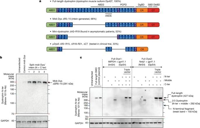
A method is developed for expressing large dystrophins to enhance muscle function in mouse models of muscular dystrophy, with potential clinical benefits for numerous disorders caused by mutations in large genes that exceed the adeno-associated virus capacity.
Category: biotech/medical – Page 717

Current biomarkers may be ineffective for evaluating heart failure risk post-pregnancy
Biomarkers used to predict heart failure risk in the general population may be ineffective for assessing risk after pregnancies complicated by hypertension or diabetes, according to a study published in JAMA Cardiology.
Several adverse pregnancy outcomes, including preeclampsia and gestational diabetes, have been linked to long-term heart health risks for pregnant women, said Priya Freaney, MD, ‘22 GME, assistant professor of Medicine in the Division of Cardiology, who was first author of the study.
“We know that features of complicated pregnancies can impact a woman’s heart disease risk decades later,” Freaney said. “It’s important for us to have some way to track and screen the patients that have pregnancy complications to further clarify if someone’s on a high-risk path toward heart disease and help bring their risk down with aggressive screening, prevention or early implementation of therapies.”

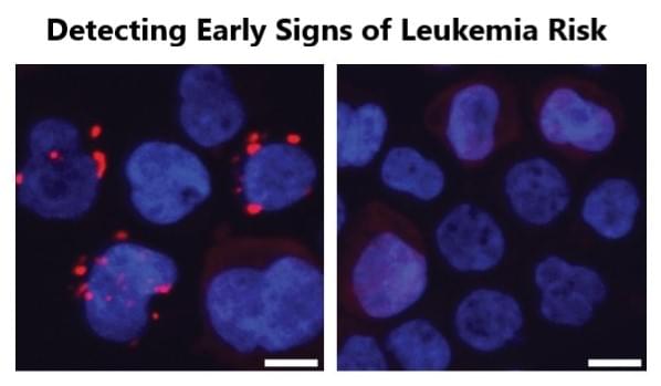
How bacteria in our aging guts can elevate risk of leukemia and perhaps more
New findings in Nature reveal how age-related gut changes fuel the growth of pre-leukemic blood cells. Scientists at Cincinnati Children’s along with an international team of researchers have discovered a surprising new connection between gut health and blood cancer risk — one that could transform how we think about aging, inflammation, and the early stages of leukemia.
As we grow older — or in some cases, when gut health is compromised by disease — changes in the intestinal lining allow certain bacteria to leak their byproducts into the bloodstream. One such molecule, produced by specific bacteria, acts as a signal that accelerates the expansion of dormant, pre-leukemic blood cells, a critical step to developing full-blown leukemia.
The team’s findings — published April 23, 2025, in the journal Nature — lay out for the first time how this process works. The study also suggests that this mechanism may reach beyond leukemia to influence risk for other diseases and among older people who share a little-known condition called clonal hematopoiesis of indeterminate potential (CHIP).
Meet Ben Lamm
ITrust Capital: Use code IMPACTGO when you sign up and fund your account to get a $100 bonus at https://impacttheory.co/iTrustCapitalMay.
Shopify: Sign up for your one-dollar-per-month trial period at https://impacttheory.co/ShopifyApr.
On this mind-bending episode of Impact Theory, Tom Bilyeu sits down with Ben Lamm, the visionary entrepreneur behind Colossal Biosciences, to explore a world that sounds straight out of science fiction—yet is rapidly becoming our reality. Together, they pull back the curtain on the groundbreaking technology making de-extinction not only possible, but increasingly practical, from resurrecting woolly mammoths and dire wolves to saving endangered species and unraveling the secrets of longevity.
Ben explains how CRISPR gene editing has unlocked the power to make precise DNA changes—editing multiple genes simultaneously, synthesizing entirely new genetic blocks, and pushing the limits of what’s possible in biology and conservation. The conversation dives deep into the technical hurdles, ethical questions, and the unexpected magic of re-engineering life itself, whether it’s creating hairier, “woolly” mice or tackling the colossal challenge of artificial wombs and universal eggs.
But this episode goes way beyond Jurassic Park fantasies. Tom and Ben debate the future of human health, gene selection through IVF, the specter of eugenics, global competition in biotechnology, and how AI will soon supercharge the pace of biological engineering. They even touch on revolutionary solutions to our plastic crisis and what it means to inspire the next generation of scientists.
Get ready to have your mind expanded. This is not just a podcast about bringing back extinct creatures—it’s a deep dive into the next frontiers of life on Earth, the technologies changing everything, and the choices we’ll face as architects of our own biology. Let’s get legendary.
00:00 Meet Ben Lamm.
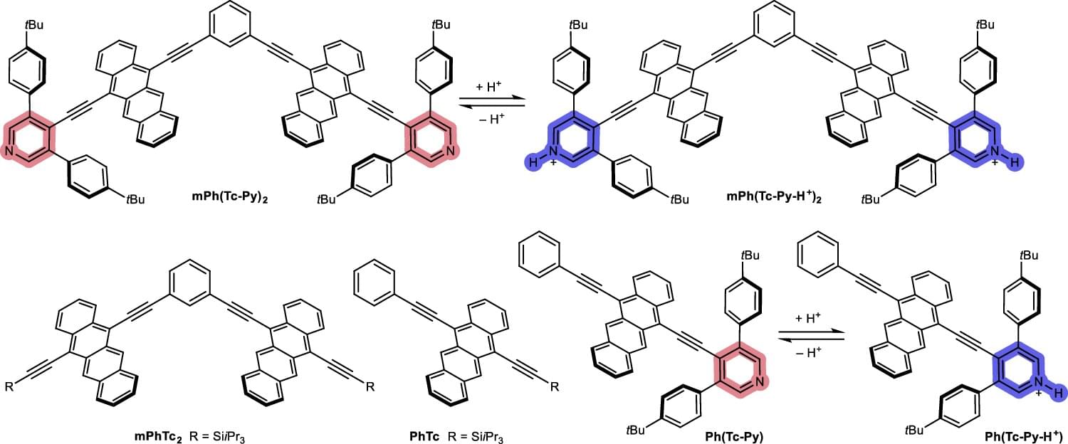
Switchable singlet fission: pH triggers molecules to split or emit light for sensors
An international team of researchers has successfully controlled the flow of energy in a molecule with the help of its pH value. The results of the study, led by Friedrich-Alexander-Universität Erlangen-Nürnberg (FAU), could contribute to the development of new sensors for medical diagnostics, for example.
The findings are also of interest for building more efficient solar cells and for use in quantum computing. The results have been published in the journal Nature Communications.
A process called singlet fission is at the center of the study. In future generations of solar cells, it should improve the utilization of light and thus increase efficiency. Until now, a large proportion of the energy that shines onto solar cells is lost and released as heat.

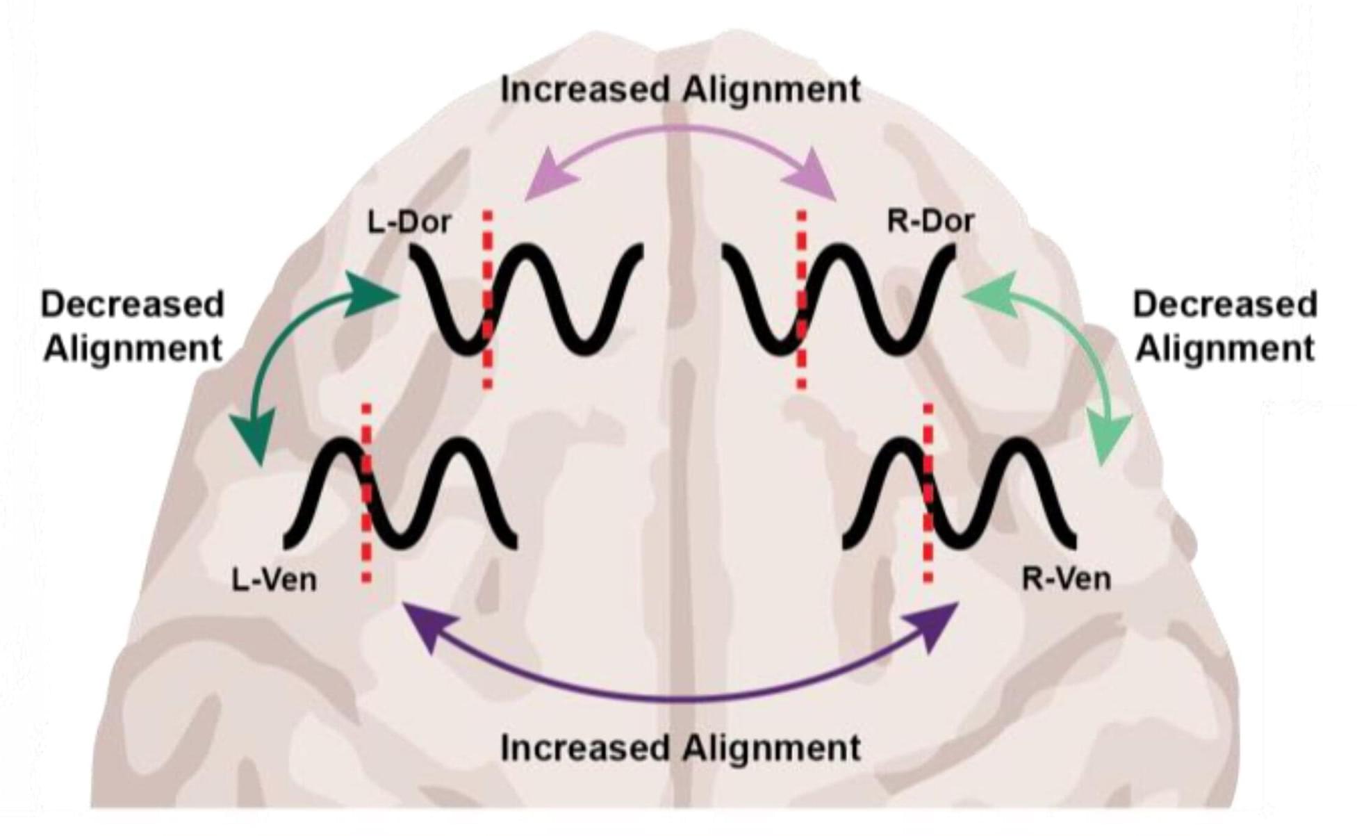
Different anesthetics, same result: Unconsciousness by shifting brainwave phase
At the level of molecules and cells, ketamine and dexmedetomidine work very differently, but in the operating room they do the same exact thing: anesthetize the patient. By demonstrating how these distinct drugs achieve the same result, a new study in animals by neuroscientists at The Picower Institute for Learning and Memory at MIT identifies a potential signature of unconsciousness that is readily measurable to improve anesthesiology care.
What the two drugs have in common, the researchers discovered, is the way they push around brain waves, which are produced by the collective electrical activity of neurons.
When brain waves are in phase, meaning the peaks and valleys of the waves are aligned, local groups of neurons in the brain’s cortex can share information to produce conscious cognitive functions such as attention, perception and reasoning, said Picower Professor Earl K. Miller, senior author of the new study in Cell Reports. When brain waves fall out of phase, local communications, and therefore functions, fall apart, producing unconsciousness.
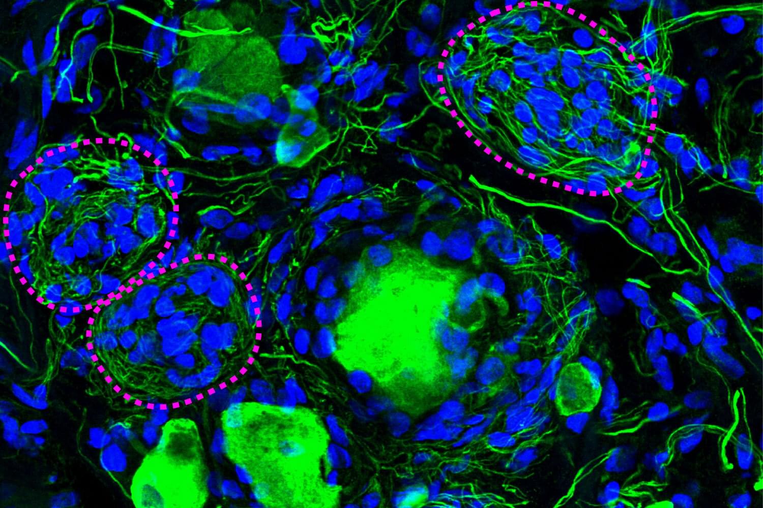
Forgotten cell clusters may hold key to diabetic neuropathy pain
A phenomenon largely ignored since its discovery 100 years ago appears to be a crucial component of diabetic pain, according to new research from The University of Texas at Dallas’s Center for Advanced Pain Studies (CAPS).
Findings from a new study published in Nature Communications suggest that cell clusters called Nageotte nodules are a strong indicator of nerve cell death in human sensory ganglia. These could prove to be a target for drugs that would protect these nerves or help manage diabetic neuropathy.
“The key finding of our study is really a new view of diabetic neuropathic pain,” said Dr. Ted Price, Ashbel Smith Professor of neuroscience in the School of Behavioral and Brain Sciences, CAPS director and co-corresponding author of the study. “We believe our data demonstrate that neurodegeneration in the dorsal root ganglion is a critical facet of the disease—which should really force us to think about the disease in a new and urgent way.”