People often compare the genome to a computer’s program, with the cell using its genetic code to process environmental inputs and produce appropriate responses.
But the machine metaphor can be extended even further to any biological system, and applying established concepts of engineering to biology could revolutionize how scientists make their observations within biology, according to research from University of Michigan.
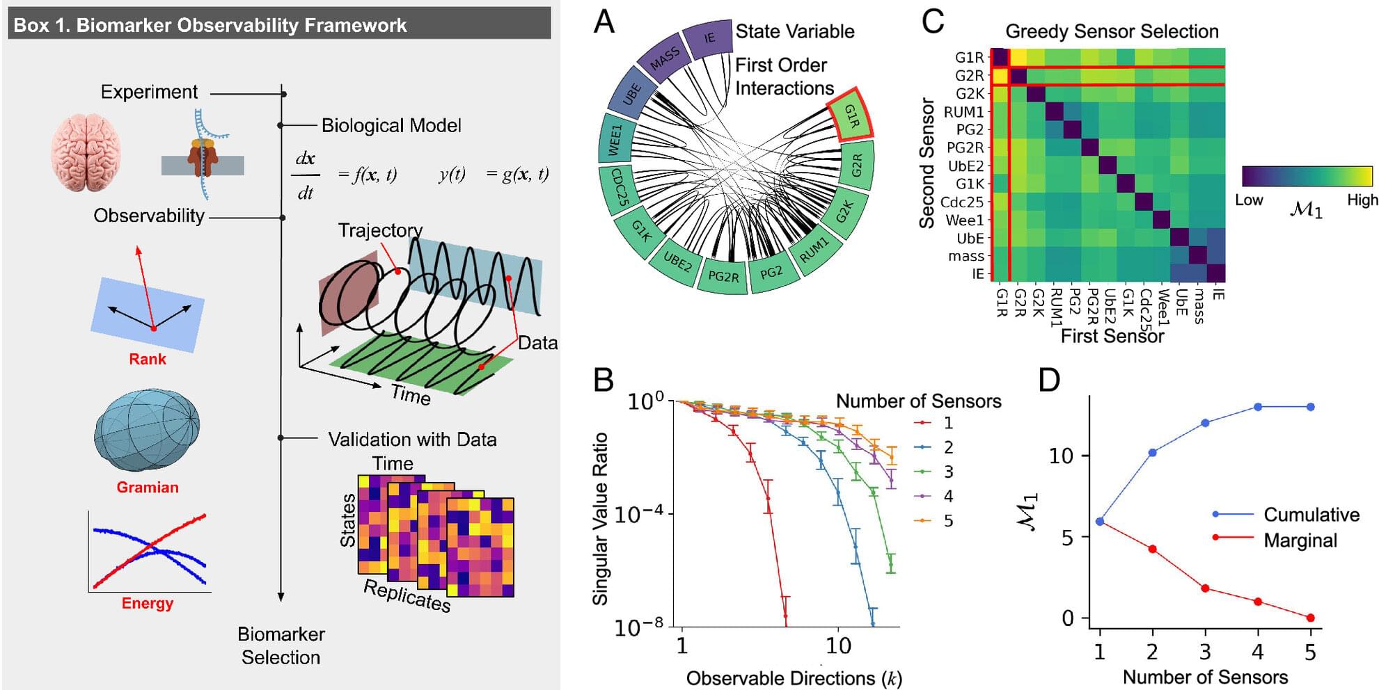
In a paper published in Proceedings of the National Academy of Sciences, Indika Rajapakse, Ph.D., Joshua Pickard, Ph.D. (now an Eric and Wendy Schmidt Postdoctoral Fellow at the Broad Institute), and their team propose that fundamental principles of control theory and observability can be applied to study biological processes that change over time.